

Idiopathic calcinosis cutis of the penis
- PMID: 23277801
- PMCID: PMC3533325
Idiopathic calcinosis cutis of the penis
Abstract
Background: Calcinosis cutis-the deposition of insoluble calcium salts in the skin and the soft tissue-occurs in the following five settings: calciphylaxis, dystrophic, iatrogenic, idiopathic, and metastatic. Idiopathic calcinosis cutis of the penis is rare.
Purpose: This paper describes a man with idiopathic calcinosis cutis of the penis, summarizes the clinical features of previously reported men with this condition, and also reviews dystrophic, iatrogenic, and metastatic penile calcinosis.
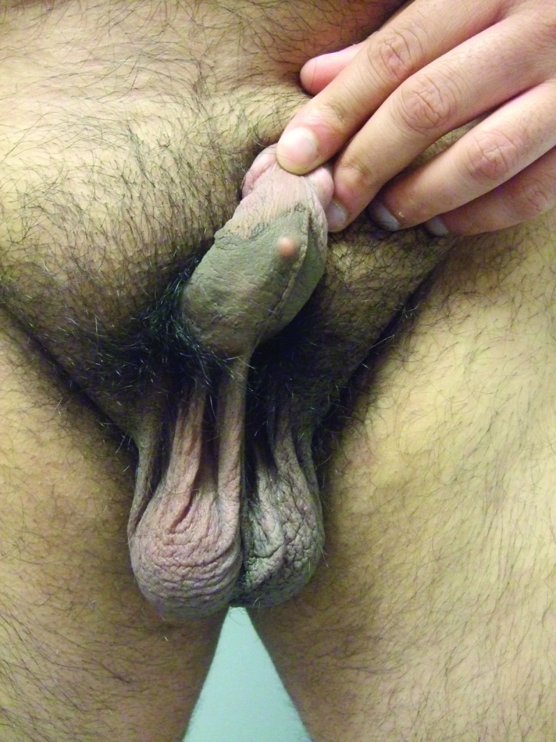
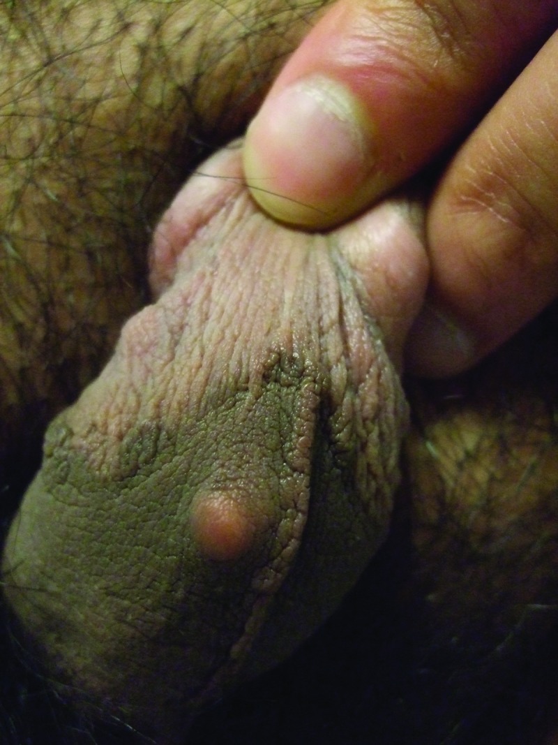
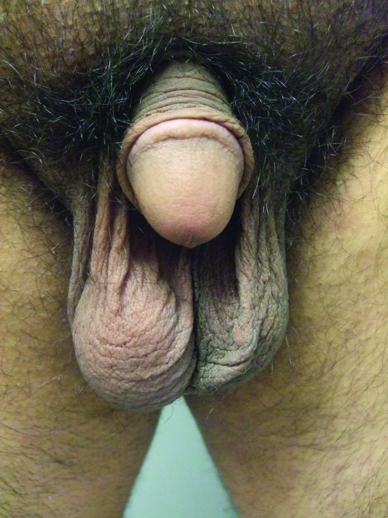
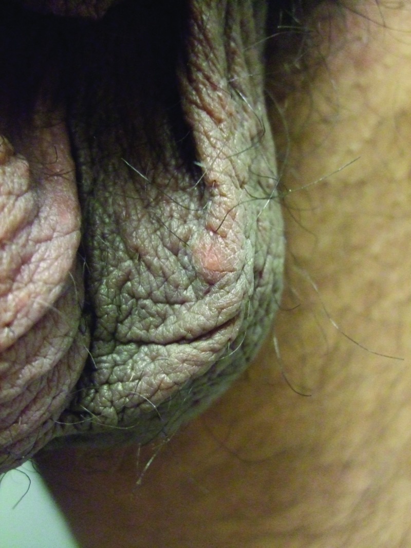
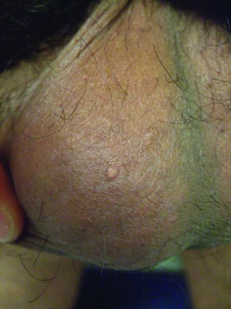
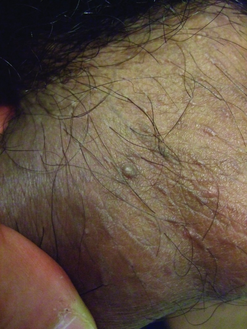
Methods: A 27-year-old Pakistan man presented with concurrent, asymptomatic, individual nodules on the right mid-ventral penile shaft and left side of scrotum and two additional papules on the right side of the scrotum. Evaluation and treatment included the excision of all lesions. Reports of patients with penile calcinosis were identified using a medical search engine (PubMed Central) and referenced citations from the published papers on this subject.
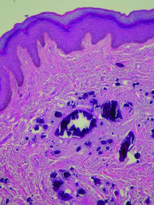
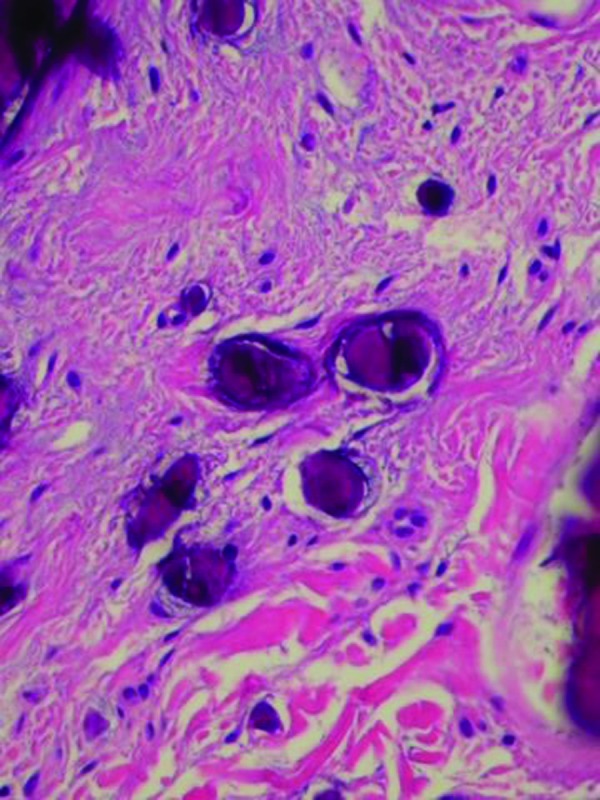
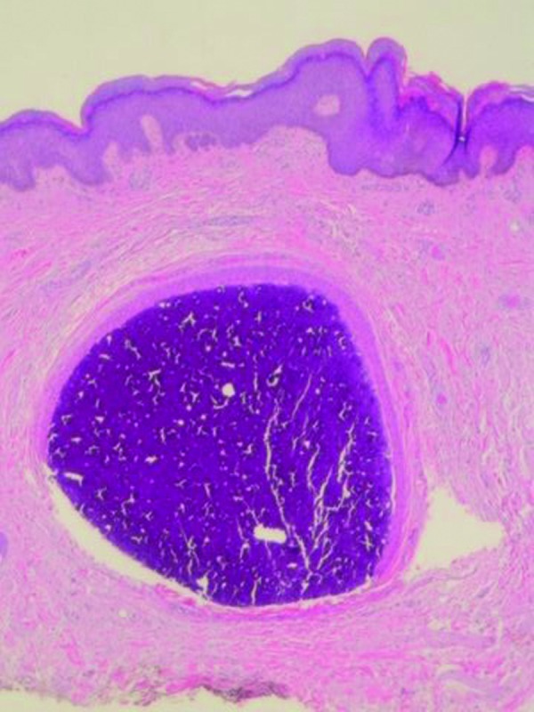
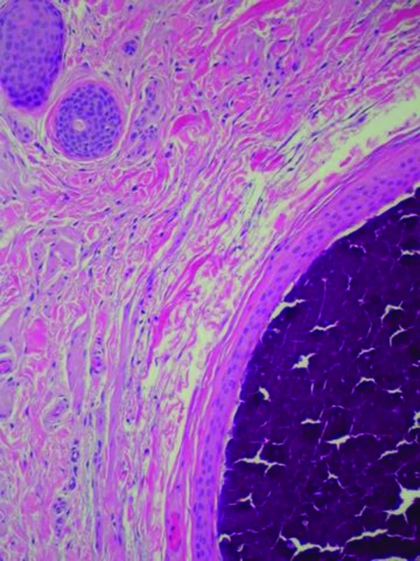
Results: Microscopic examination of the patient's nodules showed idiopathic and dystrophic calcinosis cutis of the penis and scrotum, respectively; the scrotal papules were fibroepithelial polyps. Including this individual, idiopathic calcinosis cutis of the penis has only been reported in 11 men. It presents as either an asymptomatic nodule (5 patients) or multiple lesions (6 patients) of less than one-year duration, on either the penile shaft (distal in 4 patients, mid in 2 patients, both in 1 patient, and site unspecified in 1 patient) or the prepuce (3 patients) of uncircumcised men less than 30 years of age. Concurrent scrotal calcification was noted in two patients. Dermal deposits of calcium are found in the dermis-often with surrounding histiocytes and multinucleated giant cells; concurrent features of dystrophic penile shaft calcification, such as calcium within syringomas or transepidermal elimination of calcium through eccrine sweat ducts, was only noted in two men. The nodules do not recur following excision.
Conclusion: Idiopathic calcinosis cutis of the penis is extraordinary and has only been reported in 11 men. It presents as an asymptomatic nodule or nodules on mid- to distal penile shaft or foreskin. Concurrent scrotal calcinosis cutis was noted in two men. Microscopic examination shows calcium deposits in the dermis, usually with associated histiocytes and multinucleated giant cells; concurrent changes of dystrophic calcification were also present in two men. Excision of the penile nodules not only provides the diagnosis, but also successfully resolves the condition without recurrence.
Conflict of interest statement
Figures
References
-
- Walsh JS, Fairley JA. Calcifying disorders of the skin. J Am Acad Dermatol. 1995;33:693–706. - PubMed
-
- Reiter N, El-Shabrawi L, Leinweber B, et al. Calcinosis cutis: Part I. Diagnostic pathway. J Am Acad Dermatol. 2011;65:1–12. - PubMed
-
- Dare AJ, Axelsen RA. Scrotal calcinosis: origin from dystrophic calcification of eccrine duct milia. J Cutan Pathol. 1988;15:142–149. - PubMed
-
- Ronchese F. Calcification and ossification of steatomas of the scrotum: report of a case. Arch Dermatol. 1944;49:12–15.
-
- Swinehart JM, Golitz LE. Scrotal calcinosis: dystrophic calcification of epidermoid cysts. Arch Dermatol. 1982;118:985–988. - PubMed
LinkOut - more resources
Full Text Sources