. 2012 Dec:1275:29-35.
doi: 10.1111/j.1749-6632.2012.06790.x.

Identification of DPAGT1 as a new gene in which mutations cause a congenital myasthenic syndrome

Affiliations

Identification of DPAGT1 as a new gene in which mutations cause a congenital myasthenic syndrome

Katsiaryna Belaya et al. Ann N Y Acad Sci. 2012 Dec.

Abstract

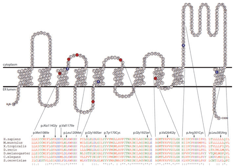
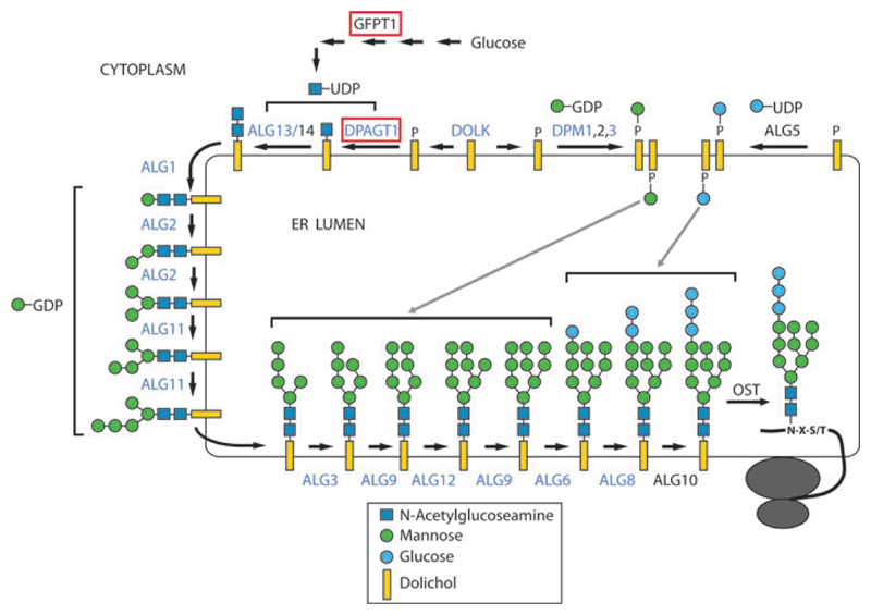
Congenital myasthenic syndromes (CMS) are a group of inherited disorders that arise from impaired signal transmission at the neuromuscular synapse. They are characterized by fatigable muscle weakness. This is a heterogenous group of disorders with 15 different genes implicated in the development of the disease. Using whole-exome sequencing we identified DPAGT1 as a new gene associated with CMS. DPAGT1 catalyses the first step of N-linked protein glycosylation. DPAGT1 patients are characterized by weakness of limb muscles, response to treatment with cholinesterase inhibitors, and the presence of tubular aggregates on muscle biopsy. We showed that DPAGT1 is required for glycosylation of acetylcholine receptor (AChR) subunits and efficient export of AChR to the cell surface. We suggest that the primary pathogenic mechanism of DPAGT1-associated CMS is reduced levels of AChRs at the endplate region. This finding demonstrates that impairment of the N-linked glycosylation pathway can lead to the development of CMS.


Conflict of interest statement

Conflicts of interest

The authors declare no conflicts of interest.

Figures

Figure 1
Figure 1
Predicted membrane topology of the DPAGT1 protein. Residues that are mutated in CMS patients are shown in red. Residues that are mutated in CDG1J patients are shown in blue. Transmembrane structure of the protein was visualized using TEXtopo. Multiple sequence alignment was performed using ClustalW.
Figure 2
Figure 2
Schematic representation of the N-linked protein glycosylation pathway. N-linked protein glycosylation takes place in the ER. It starts with the assembly of the core glycan on the lipid dolichol. Different saccharides are added to the lipid sequentially by different enzymes. The first stage of oligosaccharide assembly takes place on the cytoplasmic face of the ER, where a series of glycosyltransferases uses a cytoplasmic pool of soluble nucleotide sugar donors as substrates for dolichol glycosylation. These nucleotide-monosaccharides are synthesized by multiple cytosolic enzymes, one of which is GFPT1 (involved in the synthesis of UDP-GlcNAc). The first sugar—N-acetylglucoseamine (GlcNAc)—is added to dolichol by the enzyme DPAGT1. The second GlcNAc is transferred by the Alg13/Alg14 complex, where Alg13 is a catalytic subunit and Alg14 is an anchoring subunit that targets Alg13 to the ER membrane. Addition of the first mannose is carried out by Alg1, while Alg2 and Alg11 sequentially add four more mannose residues. Next, the resulting Dol-P-P-GlcNAc2 Man5 intermediate is flipped so that the oligosaccharide is facing inside the ER lumen. This step is carried out by an as yet unknown mechanism. Addition of subsequent sugar moieties is carried out inside the ER lumen. Here, the monosaccharide donor substrates are dolichyl-phosphate–linked monosaccharides Dol-P-mannose and Dol-P-glucose, which are synthesized on the cytoplasmic face of the ER by Dpm1/2/3 and Alg5 enzymes, respectively. Extension of Dol-P-oligosaccharide is carried out by Alg3, Alg9, and Alg12, which add four mannose residues. The final three glucose residues are added by Alg6, Alg8, and Alg10, completing the assembly of the core glycan. The core glycan is then transferred to asparagine residues of nascent proteins by the multimeric oligosaccharyl transferase complex (OST). The core glycan can then be modified inside the ER and Golgi to yield the final complex saccharide structure found on the mature proteins. Mutations in many of the enzymes of the N-linked protein glycosylation pathway have been associated with diseases. In this figure, genes in which mutations are known to lead to the development of CDG disorders are shown in blue. Genes in which mutations cause development of CMS are enclosed in red boxes.

Similar articles

Cited by

References

    1. Chaouch A, Beeson D, Hantai D, Lochmuller H. 186th ENMC international workshop: congenital myasthenic syndromes 24–26 June 2011, Naarden, The Netherlands. Neuromuscul Disord. 2012;22:566–576. - PubMed
    1. Engel AG. Current status of the congenital myasthenic syndromes. Neuromuscul Disord. 2012;22:99–111. - PMC - PubMed
    1. Senderek J, et al. Hexosamine biosynthetic pathway mutations cause neuromuscular transmission defect. Am J Hum Genet. 2011;88:162–172. - PMC - PubMed
    1. Guergueltcheva V, et al. Congenital myasthenic syndrome with tubular aggregates caused by GFPT1 mutations. J Neurol. 2012;259:838–850. - PubMed
    1. Belaya K, et al. Mutations in DPAGT1 cause a limb-girdle congenital myasthenic syndrome with tubular aggregates. Am J Hum Genet. 2012;91:193–201. - PMC - PubMed

Publication types

MeSH terms

Substances