Review
. 2013 Jan;251(1):65-79.
doi: 10.1111/imr.12017.

How the immune system talks to itself: the varied role of synapses

Affiliations
Review

How the immune system talks to itself: the varied role of synapses

Jianming Xie et al. Immunol Rev. 2013 Jan.

Abstract

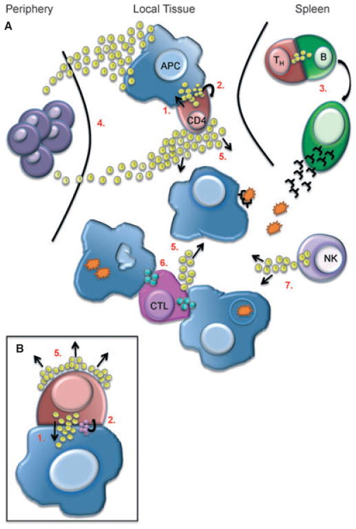
Using an elaborately evolved language of cytokines and chemokines as well as cell-cell interactions, the different components of the immune system communicate with each other and orchestrate a response (or wind one down). Immunological synapses are a key feature of the system in the ways in which they can facilitate and direct these responses. Studies analyzing the structure of an immune synapse as it forms between two cells have provided insight into how the stability and kinetics of this interaction ultimately affect the sensitivity, potency, and magnitude of a given response. Furthermore, we have gained an appreciation of how the immunological synapse provides directionality and contextual cues for downstream signaling and cellular decision-making. In this review, we discuss how using a variety of techniques, developed over the last decade, have allowed us to visualize and quantify key aspects of the dynamic synaptic interface and have furthered our understanding of their function. We describe some of the many characteristics of the immunological synapse that make it a vital part of intercellular communication and some of the questions that remain to be answered.


Conflict of interest statement

The authors have no conflicts of interest to declare.

Figures

Fig. 1
Fig. 1. Four stages of the immune synapse
In Stage 1, CD4+ T cells (red cell) extend pseudopodia causing deep invagination of the antigen-presenting cell (APC) (blue cell) cell membrane within 1 h of recognition of its cognate antigen. During Stage 2, centrioles (blue rectangles) realign themselves toward the IS and MT initiating sites (green bursts) form along the membrane that is in contact with the APC. In Stage 3, centrioles move within close proximity to the IS and the Golgi complex (yellow lines) migrates centrally to the contact site, while other organelles such as mitochondria are pushed away from it. During Stage 4, an enlarged Golgi complex is observed directly beneath the IS and the cell membrane at the T/APC contact site becomes smooth and flat.
Fig. 2
Fig. 2. A photocrosslinkable pMHC ligand revealed that ligand-bound TCRs were preferentially transported to the cSMAC
(A) A photocrosslinker azidosalicyclic acid (ASA) was introduced to a cysteine residue at P-3 position of the MCC peptide. Loading this MCC derivative to murine class II MHC I-Ek led to a photocrosslinkable pMHC, which could covalently bind to 5C.C7 TCRs on live T-cell surfaces under ultraviolet irradiation with excellent specificity and efficiency. (B) Video fluorescent imaging showed that ligand-bound TCRs were preferentially transported to the cSMAC, while free TCRs remained randomly distributed on the T-cell surface in the initial phase of T-cell activation. Ligand-bound TCRs (in green) were labeled with Alexa Fluor 555 conjugates of monovalent streptavidin (via the biotin on the associated pMHC molecules); free TCRs (in red) were labeled with Alexa Fluor 647–conjugated antigen-binding fragments of antibody KJ25 to Vβ3.
Fig. 3
Fig. 3. A photoactivatable pMHC ligand enabled the precise temporal determination of early events in TCR signaling
(A) A photoactivatable agonist peptide NPE-MCC was generated by attaching the 1-ortho-nitrophenyl-ethyl urethane (NPE) moiety to the ε-amino group of Lys99, the key TCR-recognition residue, in the MCC peptide. Irradiation with ultraviolet light would cleave off the NPE group to yield the native MCC peptide. The NPE-MCC/I–Ek complex was produced and coated to the glass surface and 5C.C7 T-cell blasts were attached to this surface. A brief UV-laser illumination would expose MCC/I–Ek in situ and enable the measurement of early events of T-cell activation with subsecond time resolution as well as micron-scale spatial resolution. (B) Summary of the timing of several key events in T-cell activation, as determined using the above technology in two studies (70, 75).
Fig. 4
Fig. 4. A real-time FRET system for analysis of TCR–pMHC interactions in situ
(A) A fluorescence resonance energy transfer (FRET) system was developed to analyze the in situ kinetic parameters of TCR binding to pMHC in synapses. TCRs on the T-cell surface were labeled with Cy3-conjugated H57-scFv, and peptide–MHCs on the planar lipid bilayer were labeled with Cy5 (at the C-terminal extension of bound peptides). The distance between Cy5 and Cy3 was about 41 Å. (B) FRET signals were observed when 5C.C7 transgenic T cells contacted the bilayer presenting agonist MCC–I-Ek ligands. (C) FRET signals were not observed when the bilayer presented null pMHC ligands.
Fig. 5
Fig. 5. Immune cell communication
The IS promotes the exchange of information by directing secretion of soluble proteins, such as cytokines (yellow spheres) or cytolytic granules (blue spheres) between two specific cells. (A) As in the case of CD4/APC interaction upon TCR recognition of a specific pMHC complex secretion is directed in a paracrine (1), as well as autocrine (2) fashion (See also inset B) which helps promote CD4 activation and polarization to a T-helper (Th) cell. Differentiated Th cells migrate to the spleen where they can promote B-cell activation and plasma cell formation, also through directed secretion of cytokine via the synapse (3). Secretion of cytokines and chemokines directed away from the IS help to recruit innate immune cells from the periphery for immediate host defense (4) or promote bystander activation during an immune response (5). Cytotoxic T lymphocytes (CTL) are able to form synapses with multiple target cells for simultaneous killing via directed secretion of cytolytic granules (6). Finally, activated immune cells, such as NK cells, can secrete cytokine in the absence of a stable IS to amplify the immune response (7).

References

    1. Schluter SF, Bernstein RM, Bernstein H, Marchalonis JJ. ‘Big Bang’ emergence of the combinatorial immune system. Dev Comp Immunol. 1999;23:107–111. - PubMed
    1. Newell EW, Sigal N, Bendall SC, Nolan GP, Davis MM. Cytometry by time-of-flight shows combinatorial cytokine expression and virus-specific cell niches within a continuum of CD8+ T cell phenotypes. Immunity. 2012;36:142–152. - PMC - PubMed
    1. Harwood NE, Batista FD. Early events in B cell activation. Annu Rev Immunol. 2010;28:185–210. - PubMed
    1. Orange JS. Formation and function of the lytic NK-cell immunological synapse. Nat Rev Immunol. 2008;8:713–725. - PMC - PubMed
    1. Monks CR, Freiberg BA, Kupfer H, Sciaky N, Kupfer A. Three-dimensional segregation of supramolecular activation clusters in T cells. Nature. 1998;395:82–86. - PubMed

Publication types