Review
. 2013 Jan;251(1):160-76.
doi: 10.1111/imr.12023.

Stromal and hematopoietic cells in secondary lymphoid organs: partners in immunity

Affiliations
Review

Stromal and hematopoietic cells in secondary lymphoid organs: partners in immunity

Deepali Malhotra et al. Immunol Rev. 2013 Jan.

Abstract

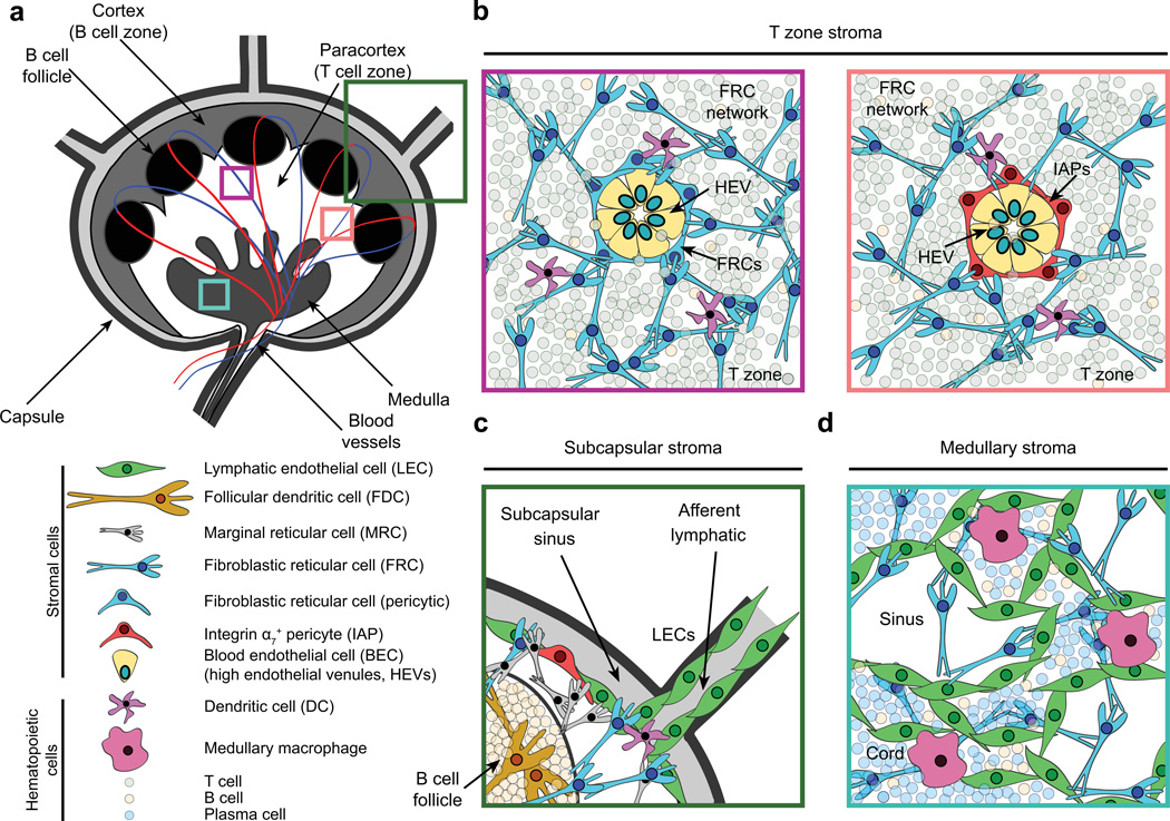
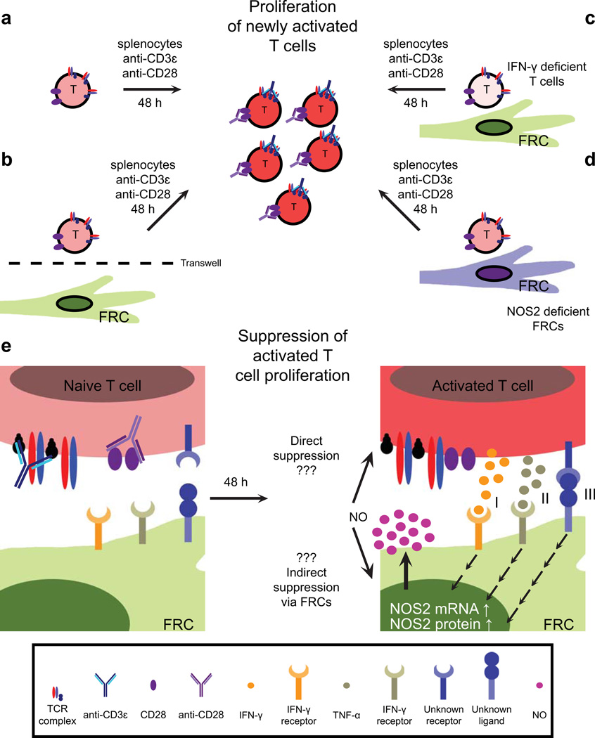
Secondary lymphoid organs (SLOs), including lymph nodes, Peyer's patches, and the spleen, have evolved to bring cells of the immune system together. In these collaborative environments, lymphocytes scan the surfaces of antigen-presenting cells for cognate antigens, while moving along stromal networks. The cell-cell interactions between stromal and hematopoietic cells in SLOs are therefore integral to the normal functioning of these tissues. Not only do stromal cells physically construct SLO architecture but they are essential for regulating hematopoietic populations within these domains. Stromal cells interact closely with lymphocytes and dendritic cells, providing scaffolds on which these cells migrate, and recruiting them into niches by secreting chemokines. Within lymph nodes, stromal cell-ensheathed conduit networks transport small antigens deep into the SLO parenchyma. More recently, stromal cells have been found to induce peripheral CD8(+) T-cell tolerance and control the extent to which newly activated T cells proliferate within lymph nodes. Thus, stromal-hematopoietic crosstalk has important consequences for regulating immune cell function within SLOs. In addition, stromal cell interactions with hematopoietic cells, other stroma, and the inflammatory milieu have profound effects on key stromal functions. Here, we examine ways in which these interactions within the lymph node environment influence the adaptive immune response.


Conflict of interest statement

The authors have no conflicts of interest to declare.

Figures

Fig. 1
Fig. 1. Lymph node architecture and stromal cell localization
(A) Cartoon depicting lymph node architecture and compartmentalization. (B) Naive lymphocytes gain access to lymph nodes through high endothelial venules (HEVs). HEVs are comprised of specialized blood endothelial cells (BECs) that regulate lymphocyte entry into lymph nodes via expression of molecules such as peripheral node addressins. These structures are routinely surrounded by a layer of fibroblastic reticular cells (FRCs, left). However, a small proportion (<15%) of these gateways are instead ensheathed by integrin α7+ pericytes (IAPs, right). Both types of HEVs appear similar histologically. Furthermore, they connect to the dense FRC network within the paracortex, providing a continuous scaffold on which naive lymphocytes and dendritic cells crawl and interact. FRCs also promote naive T-cell survival through the secretion of IL-7. (C) Interstitial fluid and migratory dendritic cells enter lymph nodes through afferent lymphatics, which are lined by lymphatic endothelial cells (LECs). Between the capsule and the lymph node parenchyma lies the subcapsular sinus (SCS), within which lymph percolates, allowing antigen uptake by macrophages. FRCs, CXCL13-secreting marginal reticular cells, and IAPs are also found proximal to the SCS. B-cell follicles contain a specialized stromal subset (follicular dendritic cells), which interacts with B lymphocytes and continually captures and displays antigens on its surface via complement receptors. (D) The lymph node medulla contains macrophages, B cells, plasma cells, and a dense LEC network that regulates lymphocyte egress through efferent lymphatics.
Fig. 2
Fig. 2. Fibroblastic reticular cell network
Fibroblastic reticular cells (FRCs) secrete, ensheath, and maintain an extracellular matrix (ECM)-based conduit or reticular network. The conduit network links the SCS to high endothelial venules (HEVs) within the lymph node paracortex, allowing small molecules such as chemokines to traffic from inflamed tissues to HEVs. Dendritic cells in close contact with the conduit network also gain rapid access to tissuederived antigens within the paracortex. In contrast to the paracortex, within B-cell follicles the FRC network is quite sparse, with little branching. (A) Confocal immunofluorescence analysis of 15 μm thick frozen skin-draining lymph node sections stained for desmin (blue, FRCs) and the ER-TR7 antigen (green, conduit network). ER-TR7 binds to an unidentified ECM component within the microfibrillar zone of the reticular network. (B) Confocal immunofluorescence analysis of 7.5 μm thick frozen skin-draining lymph node sections stained for α-smooth muscle actin (red, FRCs), the ER-TR7 antigen (blue, conduit microfibrillar zone), and collagen XIV (green, conduit core). (C) High magnification view of the conduit network. Sections were prepared as in (B), and stained for biglycan (green, collagen core) and the ER-TR7 antigen (red, conduit microfibrillar zone).
Fig. 3
Fig. 3. Regulation of activated T-cell proliferation by fibroblastic reticular cells
(A) Experimental setup: naive splenocytes were cultured in the presence of anti-CD3ε and anti-CD28 antibodies, leading to T-cell activation and proliferation. (B) When splenocytes were cultured in the presence of fibroblastic reticular cells (FRCs) with a transwell filter separating the two populations, T cells proliferated normally and little nitric oxide (NO) was detected. (C,D) Coculture and activation of T cells deficient in IFN-γ expression with wildtype (WT) FRCs, or WT T cells with FRCs deficient in NOS2 expression allows normal proliferation of T cells. (E) Activation and coculture of WT T cells with WT FRCs leads to high levels of NO in the culture supernatant, and a significant block in activated T-cell proliferation. Activated T-cell-derived IFN-γ (I), TNF-α (II), and an unknown membrane-bound signal (III) synergize to increase NOS2 mRNA and protein levels in FRCs. This in turn enhances FRC production and secretion of NO, which directly or indirectly curbs proliferation of these T cells.

References

    1. Kotturi MF, et al. Naive precursor frequencies and MHC binding rather than the degree of epitope diversity shape CD8+ T cell immunodominance. J Immunol. 2008;181:2124–2133. - PMC - PubMed
    1. Moon JJ, et al. Naive CD4(+) T cell frequency varies for different epitopes and predicts repertoire diversity and response magnitude. Immunity. 2007;27:203–213. - PMC - PubMed
    1. Obar JJ, Khanna KM, Lefrancois L. Endogenous naive CD8+ T cell precursor frequency regulates primary and memory responses to infection. Immunity. 2008;28:859–869. - PMC - PubMed
    1. Link A, et al. Fibroblastic reticular cells in lymph nodes regulate the homeostasis of naive T cells. Nat Immunol. 2007;8:1255–1265. - PubMed
    1. Malhotra D, et al. Transcriptional profiling of stroma from inflamed and resting lymph nodes defines immunological hallmarks. Nat Immunol. 2012;13:499–510. - PMC - PubMed

Publication types