

An MRI-based index to measure the severity of Alzheimer's disease-like structural pattern in subjects with mild cognitive impairment
- PMID: 23278858
- PMCID: PMC3605230
- DOI: 10.1111/joim.12028
An MRI-based index to measure the severity of Alzheimer's disease-like structural pattern in subjects with mild cognitive impairment
Abstract
Background: Structural magnetic resonance imaging (MRI) is sensitive to neurodegeneration and can be used to estimate the risk of converting to Alzheimer's disease (AD) in individuals with mild cognitive impairment (MCI). Brain changes in AD and prodromal AD involve a pattern of widespread atrophy. The use of multivariate analysis algorithms could enable the development of diagnostic tools based on structural MRI data. In this study, we investigated the possibility of combining multiple MRI features in the form of a severity index.
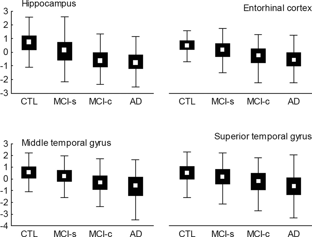
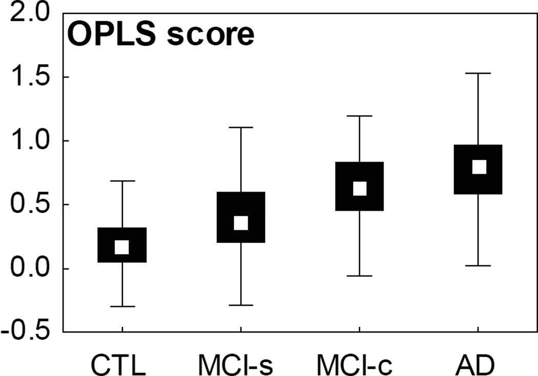
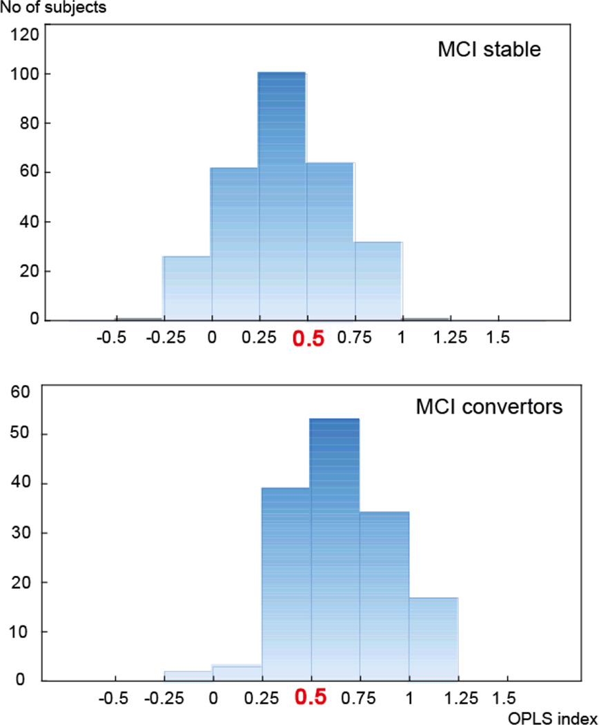
Methods: We used baseline MRI scans from two large multicentre cohorts (AddNeuroMed and ADNI). On the basis of volumetric and cortical thickness measures at baseline with AD cases and healthy control (CTL) subjects as training sets, we generated an MRI-based severity index using the method of orthogonal projection to latent structures (OPLS). The severity index tends to be close to 1 for AD patients and 0 for CTL subjects. Values above 0.5 indicate a more AD-like pattern. The index was then estimated for subjects with MCI, and the accuracy of classification was investigated.
Results: Based on the data at follow-up, 173 subjects converted to AD, of whom 112 (64.7%) were classified as AD-like and 61 (35.3%) as CTL-like.
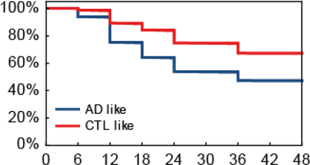
Conclusion: We found that joint evaluation of multiple brain regions provided accurate discrimination between progressive and stable MCI, with better performance than hippocampal volume alone, or a limited set of features. A major challenge is still to determine optimal cut-off points for such parameters and to compare their relative reliability.
© 2012 The Association for the Publication of the Journal of Internal Medicine.
Conflict of interest statement
No conflict of interest was declared.
Figures
References
-
- Albert MS, DeKosky ST, Dickson D, Dubois B, Feldman HH, Fox NC, Gamst A, Holtzman DM, Jagust WJ, Petersen RC, Snyder PJ, Carrillo MC, Thies B, Phelps CH. The diagnosis of mild cognitive impairment due to Alzheimer's disease: recommendations from the National Institute on Aging-Alzheimer's Association workgroups on diagnostic guidelines for Alzheimer's disease. Alzheimers Dement. 2011;7:270–279. - PMC - PubMed
-
- Mariani E, Monastero R, Mecocci P. Mild cognitive impairment: a systematic review. J Alzheimers Dis. 2007;12:23–35. - PubMed
-
- Gauthier S, Reisberg B, Zaudig M, Petersen RC, Ritchie K, Broich K, Belleville S, Brodaty H, Bennett D, Chertkow H, Cummings JL, de Leon M, Feldman H, Ganguli M, Hampel H, Scheltens P, Tierney MC, Whitehouse P, Winblad B. Mild cognitive impairment. Lancet. 2006;367:1262–1270. - PubMed
Publication types
MeSH terms
Grants and funding
LinkOut - more resources
Full Text Sources
Other Literature Sources
Medical
