. 2013 May;15(5):689-700.
doi: 10.1111/cmi.12078. Epub 2012 Dec 20.

Cell entry of Lassa virus induces tyrosine phosphorylation of dystroglycan

Affiliations

Cell entry of Lassa virus induces tyrosine phosphorylation of dystroglycan

Marie-Laurence Moraz et al. Cell Microbiol. 2013 May.

Abstract

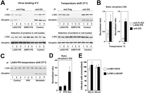
The extracellular matrix (ECM) receptor dystroglycan (DG) serves as a cellular receptor for the highly pathogenic arenavirus Lassa virus (LASV) that causes a haemorrhagic fever with high mortality in human. In the host cell, DG provides a molecular link between the ECM and the actin cytoskeleton via the adapter proteins utrophin or dystrophin. Here we investigated post-translational modifications of DG in the context of LASV cell entry. Using the tyrosine kinase inhibitor genistein, we found that tyrosine kinases are required for efficient internalization of virus particles, but not virus-receptor binding. Engagement of cellular DG by LASV envelope glycoprotein (LASV GP) in human epithelial cells induced tyrosine phosphorylation of the cytoplasmic domain of DG. LASV GP binding to DG further resulted in dissociation of the adapter protein utrophin from virus-bound DG. This virus-induced dissociation of utrophin was affected by genistein treatment, suggesting a role of receptor tyrosine phosphorylation in the process.


Figures

Fig. 1
Fig. 1. Genistein inhibits internalization of rLCMV-LASVGP
(A) LASV infection in WI-26 VA4 cells is mediated by DG. Monolayers of WI-26 VA4 cells in M96 plates were blocked with MAb IIH6 or an unrelated mouse IgM (Control IgM) at the indicated concentrations for 2 h at 4°C. Next, 200 PFU of rLCMV-LASVGP was added for 45 min. Infection was assessed after 16 hours by immunofluorescence (IF) staining for LCMV NP. Infected foci were counted in each well (means ± SD, n = 3). (B) Cytotoxicity of genistein. WI-26 VA4 cells were treated with the indicated concentrations of the drug for 4 hours, followed by a wash out and incubation for a total of 16 hours. Cell viability was assessed by Cell Titer Glo® assay. Data are triplicates ± SD. (C) Inhibition of rLCMV-LASVGP infection with genistein. WI-26 VA4 cells were treated with the indicated concentrations of genistein for one hour, followed by infection with rLCMV-LASVGP at multiplicity of 1, followed by wash out of the drug at 4 hours. After 12 hours of culture in presence of 20 mM ammonium chloride, cells were fixed and infection detected by IF for LCMV NP. Data are triplicates ± SD. (D) Inhibition of infection of VSV pseudotypes with genistein. WI-26 VA4 cells were treated with the indicated concentrations of genistein for one hour, followed by infection with rVSV-LASVGP and rVSV-VSVG (200 PFU/well). After 24 hours, infection was assessed by detection of GFP positive cells in direct fluorescence microscopy (n = 3 ± SD). (E) Schematic of the virus internalization assay (for details, please see text). (F) Genistein treatment prevents internalization of rLCMV-LASVGP. WI-26 VA4 cells were pre-treated with 50 μM of genistein or vehicle control (DMSO). After one hour, cells were chilled on ice and incubated with biotin-S-S-labeled rLCMV-LASVGP (100 particles/cell) for one hour in the cold. Unbound virus was removed, cells shifted to 37 °C in presence or absence of genistein. After the indicated time points, cells were chilled on ice and treated with TCEP (+TCEP) or reaction buffer only (−TCEP). After quenching of residual TCEP, cells were lysed, viral GP isolated by IP with mAb 83.6 to GP2. Biotinylated GP2 was detected with streptavidine-HRP in Western-blot under nonreducing conditions using enhanced chemiluminescence (ECL). The Upper blot (+TCEP) was exposed for 10 minutes, the lower blot (−TCEP) was exposed for 1 minute.
Fig. 2
Fig. 2. Binding of LASV to cellular DG induces tyrosine phosphorylation of β-DG by src-family kinases
(A) Schematic representation of C-terminally tagged DG (DGHA). The N-terminal domain (white), the mucin-type domain (black) and the C-terminal domain (gray) of α-DG, β-DG, and the C-terminal HA tag are indicated. (B). Detection of tyrosine phosphorylation at residue Y892 with mAb cl14a. DGHA was transiently expressed either alone or in combination with c-src. Parallel specimens were pretreated with 20 μM PP2 or mock treated with vehicle (DMSO). After 48 hours, DGHA was isolated by pull-down with HA matrix. Proteins were separated and probed in Western blot with an antibody to HA (anti-HA) or mAb cl14a to β-DG phosphorylated at tyrosine 892 (anti-β-DG PY892). Apparent molecular masses and the positions of β-DG are indicated. (C) Attachment of rLCMVLASVGP to cells induces tyrosine phosphorylation of β-DG. Monolayers of WI-26 VA4 cells were incubated with rLCMV-LASVGP or PICV (100 particles/cell) for 1 hour in the cold. Unbound virus was removed and cells shifted to 37°C. At the indicated time points, cells were lysed and DG enriched by WGA affinity purification. WGA-bound glycoproteins were probed in Western-blot with mAb cl14a (anti-β-DG PY892) and antibody 8D5 to β-DG. The positions of β-DG and β-DG PY892 are indicated. (D) Virus induced tyrosine phosphorylation of β-DG is blocked by PP2. Monolayers of WI-26 VA4 cells were pretreated with 20 μM PP2 or DMSO (Control) for 1 hour prior to exposure to rLCMV-LASVGP. Virus-induced phosphorylation of β-DG at Y892 was assessed as in (C). (E) The phosphorylation of β-DG at PY892 is not required for LASV cell entry. Monolayers of WI-26 VA4 cells were pretreated with 20 μM PP2 or DMSO (Control) for 1 hour as in (D), followed by incubation with rLCMV-LASVGP (MOI = 1) in the cold in presence of the drug. After 1 hour, unbound virus was removed and pre-warmed (37 °C) medium containing the drug added. At the indicated time points, 20 mM ammonium chloride was added and left throughout the experiment. At 16 hours post infection, cells were fixed and infection detected by intracellular staining for LCMV NP (means ± SD, n = 3). The apparent differences in infection at 60 minutes were not statistically significant.
Fig. 3
Fig. 3. Binding of LASV to cellular DG induces tyrosine phosphorylation of β-DG by non-src tyrosine kinases
(A) Sequence of the cytoplasic domain of human β-DG. The putative transmembrane domain is underlined in black and tyrosine residues indicated with gray arrows. Putative phosphorylation sites for src-family kinases are underlined and Y residues marked (arrows). The PPxY motif including Y892 is double underlined. (B) Detection of virus-induced tyrosine phosphorylation β-DG in presence of PP2. WI-26 VA4 cells were pretreated with 20 μM PP2 or vehicle, followed by exposure to rLCMV-LASVGP as in Fig. 2D). After the indicated time points, β-DG was isolated by IP using mAb 8D5 against β-DG covalently coupled to Sepharose 4B. Immunocomplexes were eluted under non-reducing conditions, separated by SDS-PAGE and probed in Western blot using mAb cl14a to β-DGPY892 and mAb 4G10 to tyrosine phosphate (pY). For detection of bound mouse IgG a TrueBlot® detection system was used to avoid cross-reaction with the murine IgG heavy and light chains. Total β-DG was detected with rabbit polyclonal antibody AP83. (C) Genistein blocks virus-induced tyrosine phosphorylation of β-DG. Cells were pre-treated with 50 μM genistein or vehicle (DMSO) only. After 30 minutes, rLCMV-LASVGP was added (100 particles/cell) for the indicated time points and tyrosine phosphorylation of β-DG was assessed as in (B).
Fig. 4
Fig. 4. Retroviral pseudotypes
(A) Schematic representation of Flag-tagged LASV GP. The receptor-binding GP1 and transmembrane GP2 parts are indicated. The transmembrane domain of GP2 is represented as a grey box and the C-terminal FLAG-tag indicated. (B) Detection of GP in retroviral pseudotypes. Retroviral pseudotypes were generated by cotransfection of packaging cell line GP2293 expressing retroviral Gag/Pol with a GP expression plasmid and an expression plasmid for a packable retroviral genome bearing a luciferase and a GFP reporter gene. Pseudotypes were purified by ultracentrifugation through a sucrose cushion and re-suspended in HBSS. Purified pseudotypes of LASV (LAS), AMPV (AMP), and VSV, as well as pseudotypes lacking GP (C) were immobilized in microtiter plates and the viral GP detected with mAb 83.6, combined with an HRP-conjugated secondary antibody in a colour reaction (means ± SD, n = 3). (C) Infection of cells with LASV pseudotypes depends on DG. DG (−/−) murine embryonic stem (ES) cells and parental DG (+/−) ES cells were infected with pseudotypes of LASV (LAS), AMPV (AMP), and VSV, as well as pseudotypes lacking GP (C). After 48 hours, infection was detected by luciferase assay (means ± SD, n = 3).
Fig. 5
Fig. 5. Effect of LASV pseudotype binding on the association of DG with utrophin
Monolayers of WI-26 VA4 cells were chilled on ice and incubated with either LASV or AMPV pseudotypes (LASV-PS, AMPV-PS) at a multiplicity of infection (MOI) of 50 transforming units (TU)/cell. Parallel specimens were incubated with mAb 16G4 to α-DG (anti-DG). After one hour, unbound virus or mAb were removed by washing. Cells were either kept on ice (virus binding 4°C) or shifted to 37 °C for 10 minutes (temperature shift 37°C). Cells were quickly chilled on ice, lysed and subjected to IP using FLAG matrix or protein G-conjugated Sepharose 4B. Immunocomplexes were separated by SDS-PAGE using 100% of the IPs anti-FLAG and 5% of the IP anti-DG. Beta-DG and utrophin were detected on Western-blot using monoclonal antibodies 8D5 and combined with HRP-conjugated secondary antibodies in a TrueBlot® detection system to avoid cross-reaction with the IgG heavy chain. For the detection of total protein in cell lysates, 1/20 of the lysate were separated by SDS-PAGE and subjected to Western-blot detection. (B) Quantification of the signals in (A). Blots were scanned in a densitometer and the ratios of the signals for utrophin normalized to β-DG (utrophin/β-DG) for the IPs anti-FLAG (LASV pseudotypes only) and the IP anti-DG. For each series, the utrophin/β-DG ratio detected in the IP anti-DG was defined as 1.0. (C) Pre-treatment with genistein, but not PP2 reduced virus-induced dissociation of utrophin from DG. Monolayers of WI-26 VA4 cells were pre-treated with DMSO only (control), 20 μM PP2 and 50 μM genistein for one hour. Cells were then chilled on ice and incubated with LASV pseudotypes (LASV-PS) for one hour in the cold in presence of drugs. Cells were then quickly shifted to 37 °C, lysed, and subjected to IP with FLAG matrix as in (A). Precipitated β-DG and utrophin were detected in Western blot and the ratios utrophin/β-DG determined as in (B). (D) Quantification of the data in (C). (E) The 15 C-terminal amino acids of β-DG are dispensable for LASV cell entry. Murine ES cells expressing either wild-type DG (DG wt) or DG lacking the C-terminal 15 amino acids of β-DG (DGΔC) were infected with rLCMV-LASVGP or rLCMV-VSVG at a multiplicity of 0.1. Infection of the cells expressing wild-type DG was set at 100% (means ± SD, n = 3).

Similar articles

Cited by

References

    1. Barresi R, Campbell KP. Dystroglycan: from biosynthesis to pathogenesis of human disease. J Cell Sci. 2006;119:199–207. - PubMed
    1. Belkin AM, Smalheiser NR. Localization of cranin (dystroglycan) at sites of cell-matrix and cell-cell contact: recruitment to focal adhesions is dependent upon extracellular ligands. Cell Adhes Commun. 1996;4:281–296. - PubMed
    1. Beyer WR, Popplau D, Garten W, von Laer D, Lenz O. Endoproteolytic processing of the lymphocytic choriomeningitis virus glycoprotein by the subtilase SKI-1/S1P. J Virol. 2003;77:2866–2872. - PMC - PubMed
    1. Borrow P, Oldstone MB. Characterization of lymphocytic choriomeningitis virus-binding protein(s): a candidate cellular receptor for the virus. J Virol. 1992;66:7270–7281. - PMC - PubMed
    1. Borrow P, Oldstone MB. Mechanism of lymphocytic choriomeningitis virus entry into cells. Virology. 1994;198:1–9. - PubMed

Publication types

MeSH terms