. 2013 Apr;12(2):319-23.
doi: 10.1111/acel.12041. Epub 2013 Jan 4.

The gastrointestinal manifestations of telomere-mediated disease

Affiliations

The gastrointestinal manifestations of telomere-mediated disease

Naudia L Jonassaint et al. Aging Cell. 2013 Apr.

Abstract

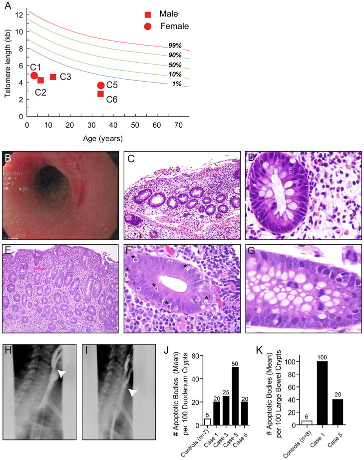
Defects in telomere maintenance genes cause pathological telomere shortening, and manifest in syndromes which have prominent phenotypes in tissues of high turnover: the skin and bone marrow. Because the gastrointestinal (GI) epithelium is highly proliferative, we sought to determine whether telomere syndromes cause GI disease, and to define its prevalence, spectrum, and natural history. We queried subjects in the Johns Hopkins Telomere Syndrome Registry for evidence of luminal GI disease. In sixteen percent of Registry subjects (6 of 38), there was a history of significant GI pathology, and 43 additional cases were identified in the literature. Esophageal stenosis, enteropathy, and enterocolitis were the recurrent findings. In the intestinal mucosa, there was striking villous atrophy, extensive apoptosis, and anaphase bridging pointing to regenerative defects in the epithelial compartment. GI disease was often the first and most severe manifestation of telomere disease in young children. These findings indicate that telomere dysfunction disrupts the epithelial integrity in the human GI tract manifesting in recognizable disease processes. A high index of suspicion should facilitate diagnosis and management.


Conflict of interest statement

Potential Competing Interests

Dr. Califano is the Director of Research of the Milton J. Dance Head and Neck Endowment. The terms of this arrangement are being managed by the Johns Hopkins University in accordance with its conflict of interest policies. The other authors have no relevant financial conflict of interest to declare.

Figures

Figure 1
Figure 1. Clinicopathologic findings in individuals with telomere-mediated gastrointestinal disease
(A) Lymphocyte telomere length is plotted relative to distribution of length in 400 controls. C1, C2....etc. refer to cases in Table 1. (B) Rectal endoscopy shows marked erythema with linear ulcerations and narrowing at the rectosigmoid junction (Case 1). (C) Colonic mucosa from Case 1 shows an expanded lamina propria with lymphoid cells and several crypts are completely absent or damaged resulting in mucosal atrophy. The mucosal surface itself is damaged and is partially sloughed off (original magnification, 20X). (D) Higher magnification of the colonic mucosa from Case 1 showing that although the lamina propria on the right side of the field is expanded with lymphoid cells, plasma cells are wholly absent, a finding typically associated with congenital immunodeficiency. The colonic crypt at the left side of the field shows an intraepithleial neutrophil infiltrate (focal acute colitis) at the 2:00 position in the crypt (original magnification 100X). (E) Although the mucosa in this image has the appearance of colonic mucosa, this biopsy was from the duodenum and shows villous atrophy and an expanded lamina propria containing lymphoplasmacytic cells (Case 5, original magnification, 20X). (F) High magnification of colonic mucosa displaying striking crypt apoptosis with significant intraepithelial lymphocytosis (Case 5, original magnification 100X). Examples of apoptotic bodies are indicated by *. (G) High magnification of colon demonstrating an anaphase bridge at the center left portion of the field (Case 5, original magnification, 125X). Inset of the anaphase bridging is shown in the right upper corner. (H) & (I) Still images from cineesophagopharyngogram demonstrating a tapered luminal defect in the cervical esophagus (Case 6). (J) & (K) Number of apoptotic bodies per 100 crypts in the duodenum and colon, respectively is plotted relative to controls.

References

    1. Addison M, Rice MS. The Association of Dyskeratosis Congenita and Fanconi’s Anaemia. Med J Aust. 1965;1:797–799. - PubMed
    1. Alter BP, Giri N, Savage SA, Rosenberg PS. Cancer in dyskeratosis congenita. Blood. 2009;113:6549–6557. - PMC - PubMed
    1. Arca E, Tuzun A, Tastan HB, Akar A, Kose O. Dyskeratosis congenita with esophageal and anal stricture. Int J Dermatol. 2003;42:555–557. - PubMed
    1. Armanios M. Syndromes of telomere shortening. Annu Rev Genomics Hum Genet. 2009;10:45–61. - PMC - PubMed
    1. Armanios M, Alder JK, Parry EM, Karim B, Strong MA, Greider CW. Short telomeres are sufficient to cause the degenerative defects associated with aging. Am J Hum Genet. 2009;85:823–832. - PMC - PubMed

Publication types

MeSH terms

LinkOut - more resources