. 2012 Dec 27;27(4):e305-13.
doi: 10.5301/JBM.2012.10437.

Thrombin stimulation of inflammatory breast cancer cells leads to aggressiveness via the EGFR-PAR1-Pak1 pathway

Affiliations

Thrombin stimulation of inflammatory breast cancer cells leads to aggressiveness via the EGFR-PAR1-Pak1 pathway

Kazufumi Ohshiro et al. Int J Biol Markers. .

Abstract

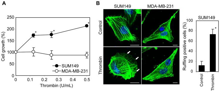
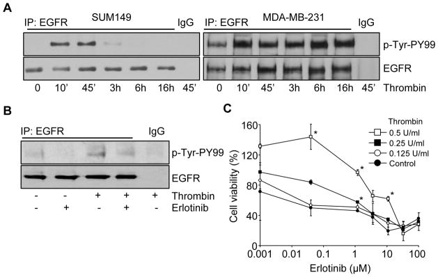
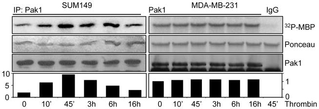
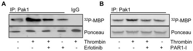
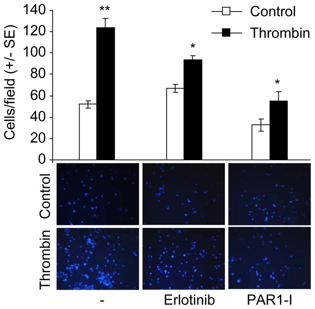
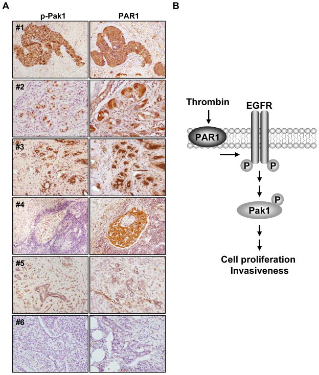
Inflammatory breast cancer (IBC) accounts for a small fraction but aggressive form of epithelial breast cancer. Although the role of thrombin in cancer is beginning to be unfolded, its impact on the biology of IBC remains unknown. The purpose of this study was to establish the role of thrombin on the invasiveness of IBC cells. The IBC SUM149 cell line was treated with thrombin in the absence or presence of the epidermal growth factor receptor (EGFR) inhibitor erlotinib and protease-activated receptor 1 (PAR1) inhibitor. The effects of pharmacological inhibitors on the ability of thrombin to stimulate the growth rate and invasiveness were examined. We found that the inhibition of putative cellular targets of thrombin action suppresses both the growth and invasiveness of SUM149 cells in a concentration-dependent manner. In addition, thrombin-mediated increased invasion of SUM149 cells was routed through EGFR phosphorylation, and in turn, stimulation of the p21-activated kinase (Pak1) activity in a EGFR-sensitive manner. Interestingly, thrombin-mediated activation of the Pak1 pathway stimulation was blocked by erlotinib and PAR1 inhibitor. For proof-of-principle studies, we found immunohistochemical evidence of Pak1 activation as well as expression of PAR1 in IBC. Thrombin utilizes EGFR to relay signals promoting SUM149 cell growth and invasion via the Pak1 pathway. The study provides the rationale for future therapeutic approaches in mitigating the invasive nature of IBC by targeting Pak1 and/or EGFR.


Figures

Figure 1
Figure 1
Thrombin stimulation of SUM149 cell growth and membrane ruffling. (A) Effect of thrombin treatment (0–0.5 U/mL) on the SUM149 and MDA-MB-231 cell growth. (B) SUM149 and MDA-MB-231 cells were maintained for 24 hours in serum-free medium and then treated with thrombin (0.5 U/mL) for 24 hours. The cells were fixed and immunofluorescent labeled with fluorescently conjugated phalloidin (for F-actin) and DAPI (for DNA). Arrows show the membrane ruffling induced by thrombin treatment. The number of SUM149 cells with the membrane ruffling were counted after taking pictures by confocal microscopy and represented as percentage of total cell number in the pictures. *P = 2.09 × 10⁻¹⁰. Scale bar = 10μm.
Figure 2
Figure 2
Thrombin-induced phosphorylation of EGFR in SUM149 cells. (A) Serum-starved SUM149 and MDA-MB-231 cells were treated with or without thrombin (0.5 U/mL) for various times before preparing the cell lysates. After immunoprecipitation of the cell lysates with anti-EGFR antibody or IgG, EGFR phosphorylation was analyzed by immunoblotting using anti-p-Tyr-PY99 antibody. The level of total EGFR was checked by immunoblotting using anti-EGFR antibody. (B) Serum-starved SUM149 cells were treated with thrombin (0.5 U/mL) and with or without Erlotinib pretreatment (50 μM), and EGFR phosphorylation was analyzed as above. (C) Cell viabilities were measured after treatment with thrombin (0–0.5 U/mL) and Erlotinib (0–100 μM). Data represent the mean ± SE. *P < 0.001.
Figure 3
Figure 3
Thrombin stimulates Pak1 activity in SUM149 cells. Cell lysates were made from SUM149 and MDA-MB-231 cells treated with thrombin (0.5 U/mL) for various times. After immunoprecipitation of the cell lysates with anti-Pak1 antibody or IgG, Pak1 kinase activity was analyzed by kinase assay using MBP as a substrate. The middle panels are Ponceau S-stained blots showing MBP. The lower panels are the total Pak1 level determined by immunoblotting using anti-Pak1 antibody.
Figure 4
Figure 4
Inhibition of thrombin-induced Pak1 activity by EGFR and PAR1 inhibitors in SUM149 cells. (A) Serum started SUM149 cells were treated with thrombin (0.5 U/mL) and with or without Erlotinib pretreatment (50 μM). After immunoprecipitation of the cell lysates with anti-Pak1 antibody or IgG, Pak1 kinase activity was analyzed by kinase assay using MBP as a substrate. The lower panels are Ponceau S-stained blots showing MBP. (B) Serum starved SUM149 cells were treated with thrombin (0.5 U/mL) and with or without PAR1-I pretreatment (500 nM), followed by cell lysis to measure the Pak1 kinase activity as above. The lower panel is Ponceau S-stained blot showing MBP.
Figure 5
Figure 5
Thrombin-induced invasiveness of the SUM149 cells. SUM149 cells (1 × 10⁵ cells) treated with or without thrombin (0.5 U/mL), Erlotinib (50 μM), or PAR1-I (200 nM) were added on upper side of matrigel invasion chamber and incubated overnight. Subsequently, the invaded cells were fixed, stained with DAPI and counted under microscope. Data represent the mean ± SE. *P < 0.01. **P < 0.001.
Figure 6
Figure 6
Status of activated p-Pak1 and PAR1 in IBC tissues (A) The expression level of p-Pak1 and PAR1 were assayed in tumor tissue samples from IBC patients using anti-p-Pak1 and anti-PAR1 antibodies. Histological characteristics of tumors #1 and #4, lymph node; #2, #3 and #5, breast tissues; #6, negative staining in lymph node. (B) Proposed working model for the activation of thrombin-induced signaling pathway via EGFR and Pak1 activation. Thrombin induces EGFR phosphorylation, and subsequently, Pak1 activation, leading to the activation of oncogenic signaling including cell proliferation and invasiveness in inflammatory breast cancer.

References

    1. Even-Ram S, Uziely B, Cohen P, Grisaru-Granovsky S, Maoz M, Ginzburg Y, et al. Thrombin receptor overexpression in malignant and physiological invasion processes. Nat Med. 1998;4:909–14. - PubMed
    1. Nierodzik ML, Karpatkin S. Thrombin induces tumor growth, metastasis, and angiogenesis: Evidence for a thrombin-regulated dormant tumor phenotype. Cancer Cell. 2006;10:355–62. - PubMed
    1. Snyder KM, Kessler CM. The pivotal role of thrombin in cancer biology and tumorigenesis. Semin Thromb Hemost. 2008;34:734–41. - PubMed
    1. Kleer CG, van Golen KL, Merajver SD. Molecular biology of breast cancer metastasis. Inflammatory breast cancer: clinical syndrome and molecular determinants. Breast Cancer Res. 2000;2:423–9. - PMC - PubMed
    1. Sportès C, Steinberg SM, Liewehr DJ, Gea-Banacloche J, Danforth DN, Avila DN, et al. Strategies to improve long-term outcome in stage IIIB inflammatory breast cancer: multimodality treatment including dose-intensive induction and high-dose chemotherapy. Biol Blood Marrow Transplant. 2009;15:963–70. - PMC - PubMed

Publication types

MeSH terms