. 2013 Apr 1;119(7):1321-9.
doi: 10.1002/cncr.27910. Epub 2012 Dec 20.

Inhibition of farnesoid X receptor controls esophageal cancer cell growth in vitro and in nude mouse xenografts

Affiliations

Inhibition of farnesoid X receptor controls esophageal cancer cell growth in vitro and in nude mouse xenografts

Baoxiang Guan et al. Cancer. .

Abstract

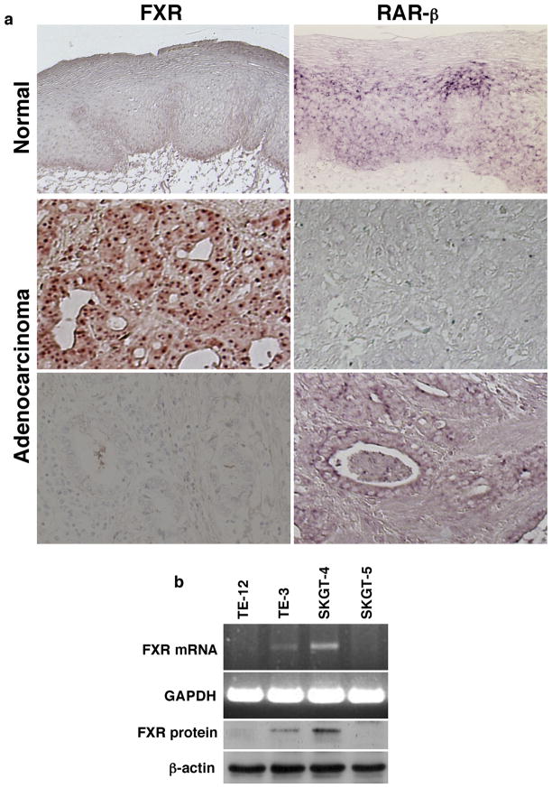
Background: Gastroesophageal reflux is a risk factor for esophageal adenocarcinoma, and bile acid and its farnesoid X receptor (FXR) have been implicated in esophageal tumorigenesis. The authors investigated the role of FXR expression and activity in esophageal cancer initiation and growth.

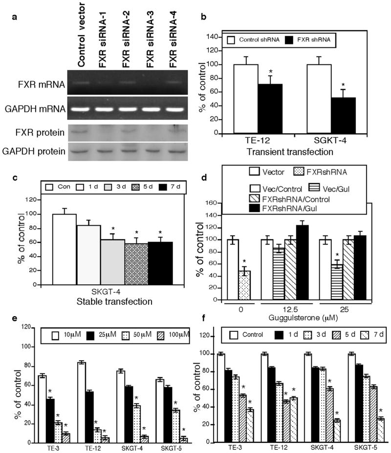
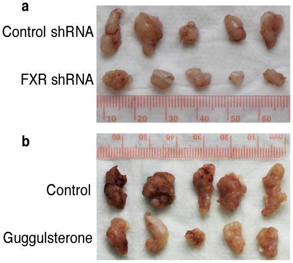
Methods: FXR expression in esophageal adenocarcinoma tissues was assessed by immunohistochemistry. Knockdown of FXR expression in esophageal cancer cells in vitro and in nude mice xenografts was suppressed by FXR small hairpin RNA (shRNA) and guggulsterone (a natural FXR inhibitor). Esophageal cancer cells were treated with bile acids to demonstrate their effects on growth-promoting genes.

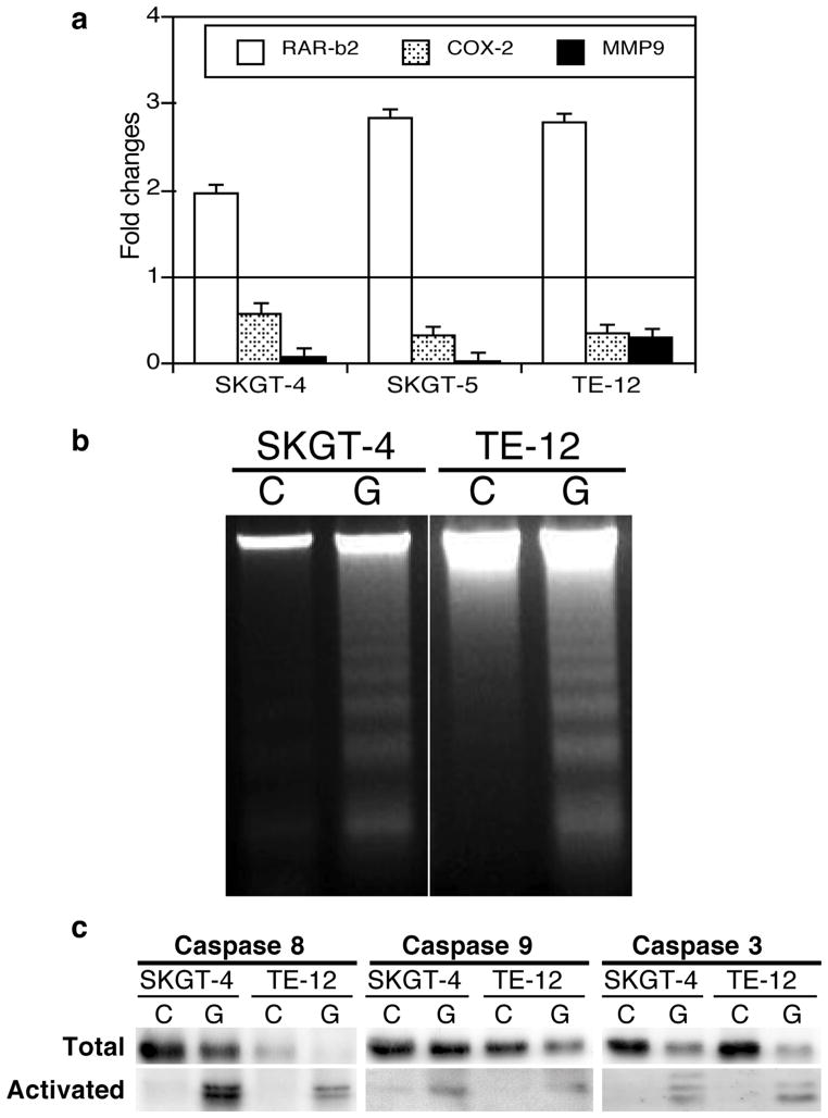
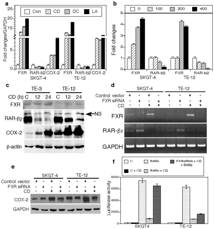
Results: FXR was expressed in 48 of 59 esophageal adenocarcinoma tissues (81.3%), and this overexpression was associated with higher tumor grade, larger tumor size, and lymph node metastasis; however, was inversely associated with retinoic acid receptor-β2 (RAR-β2 ) expression. Knockdown of FXR expression suppressed tumor cell growth in vitro and in nude mouse xenografts. Guggulsterone reduced the viability of esophageal cancer cells in a time-dependent and dose-dependent manner, whereas this effect was diminished after knockdown of FXR expression. Guggulsterone induced apoptosis through activation of caspase-8, caspase-9, and caspase-3 in tumor cells. FXR mediated bile acid-induced alterations of gene expression, eg, RAR-β2 and cyclooxygenase-2 (COX-2).

Conclusions: Inhibition of FXR by FXR shRNA or guggulsterone suppressed tumor cell viability and induced apoptosis in vitro, and it reduced tumor formation and growth in nude mouse xenografts. FXR also mediated bile acid-induced alterations of cell growth-related genes in esophageal cancer cells.


Conflict of interest statement

The authors have declared no competing interests.

Figures

Figure 1
Figure 1
Expression of FXR in esophageal cancer cells and tissue specimens. (a) Tissue samples from 59 esophageal adenocarcinoma patients were immunostained for FXR and hybridized in situ for RAR-β2 expression. The association of the expression of FXR with RAR-β2 was determined by the McNemar test. (b) Expression of FXR mRNA and protein in esophageal squamous cell carcinoma TE-3 and Te-12 cells and adenocarcinoma SKGT-4 and SKGT-5 cells using semi-quantitative RT-PCR and Western blot, respectively.
Figure 2
Figure 2
Suppression of FXR expression or activity reduces growth and proliferation of esophageal cancer cells. (a) FXR expression in SKGT-4 cells was knocked down by one of four FXR shRNA constructs. (b) Ki-67 expression was detected by immunocytochemical staining in FXR shRNA-transfected esophageal cancer cell lines. *P <0.05 compared with control cells. (c) MTT assay. SKGT-4 cells stably transfected with FXR shRNA or negative control shRNA were subjected to cell viability MTT assay. *P <0.05 compared with control cells. (d) MTT assay. SKGT-4 cells stably transfected with control shRNA (Vector or Vec) or FXR shRNA were treated or not treated (Control) with 12.5 or 25 μM guggulsterone (Gul) for 5 days and subjected to MTT assay. The data showed that FXR knockdown antagonized the effects of FXR inhibitor guggulsterone on esophageal cancer cells. *P <0.05 compared with control cells. (e) MTT assay. TE-3, TE-12, SKGT-4, and SKGT-5 cells were grown and treated with different concentrations of guggulsterone for 5 days and then subjected to MTT assay. *P <0.05 compared with control cells. (f) MTT assay. TE-3, TE-12, SKGT-4, and SKGT-5 cells were grown and treated with 25 μM guggulsterone for up to 7 days and subjected to MTT assay. *P <0.05 compared with control cells.
Figure 3
Figure 3
The FXR inhibitor guggulsterone regulates gene expression and promotes apoptosis in esophageal cancer cells. (a) qRT-PCR. Esophageal cancer SGKT-4, SGKT-5, and TE-12 cell lines were grown and treated with 25 μM guggulsterone for 2 days for qRT-PCR. Bars below the horizontal 1-fold line indicate reduced expression; those above the line, increased expression induced by guggulsterone. (b) DNA fragmentation assay. SKGT-4 and TE-12 cells were grown and treated with 25 μM guggulsterone (G) or control (C) for 3 days and then subjected to the DNA fragmentation assay to measure apoptosis. (c) Western blot. Esophageal cancer SKGT-4 and TE-12 cells were grown and treated with 25 μM guggulsterone (G) or control (C) for 3 days, and total cellular protein was extracted and subjected to Western blotting.
Figure 4
Figure 4
FXR inhibition reduces growth of esophageal cancer cells in vivo. (a) SKGT-4 cells stably transfected with control shRNA or FXR shRNA were subcutaneously injected into nude mice. Tumor formation and growth were monitored daily. (b) Nude mice were treated with 50 mg/kg of guggulsterone orally for 2 days and then subcutaneously injected with SKGT-4 cells and continued to receive 50 mg/kg of guggulsterone daily for additional 20 days. Tumor formation and growth were monitored daily. At the end experiments, tumor xenografts were taken out, weighted, and photographed.
Figure 5
Figure 5
FXR mediates the effects of bile acids on regulation of gene expression in esophageal cancer cells. (a) qRT-PCR. SKGT-4 and TE-12 cells were grown and treated with 200 μM of chenodeoxycholic acid (CD), deoxycholic acid (DC), lithocholic acid (LA), or control (Con) for 48 h for qRT-PCR. (b) qRT-PCR. SKGT-4 and TE-12 cells were grown and treated with different doses of chenodeoxycholic acid (all, μM) for qRT-PCR. (c) Western blot. Cells treated with chenodeoxycholic acid or control (C) for 12 or 24 h for Western blotting. NS, nonspecific. (d) semiquantitative RT-PCR. SKGT-4 and TE-12 cells were grown and transiently transfected with FXR shRNA-3 and treated with 200 μM chenodeoxycholic acid for 24 h for semiquantitative RT-PCR. (e) Western blot. Cells treated as for (d) were subjected to Western blotting. (f) Luciferase assay. SKGT-4 and TE-12 cells were grown and transiently transfected with RAR-β2 gene promoter-driven luciferase reporter vector with or without FXR shRNA vector or empty vector. pCH110, a β-galactosidase expression vector, was used as an internal control for assessing transfection efficiency. Thirty-six hours after transfection, the cells were treated with chenodeoxycholic acid or left untreated for an additional 24 h. The cells were then harvested, and luciferase activities were measured. The data showed that luciferase activity was high after RAR-β2 promoter transfection, while CD treatment suppressed RAR-β2 luciferase activity. However, after co-transfection with FXR shRNA, RAR-β2 luciferase activity was significantly rescued in SKGT-4 cells compared to the CD treated cells but only faintly rescued in TE-12 cells (FXR expression in TE-12 cells is very low compared to SKGT-4 cells). C, negative control; CD, chenodeoxycholic acid.

References

    1. Blot W. Esophageal cancer trends and risk factors. Semin Oncol. 1994;21:403–410. - PubMed
    1. Spechler SJ. Barrett’s esophagus: a molecular perspective. Curr Gastroenterol Rep. 2005;7:177–181. - PubMed
    1. Chen X, Yang CS. Esophageal adenocarcinoma: a review and perspectives on the mechanism of carcinogenesis and chemoprevention. Carcinogenesis. 2001;22:1119–1129. - PubMed
    1. Barak N, Ehrenpreis ED, Harrison JR, Sitrin MD. Gastro-oesophageal reflux disease in obesity: pathophysiological and therapeutic considerations. Obes Rev. 2002;3:9–15. - PubMed
    1. Makishima M, Okamoto AY, Repa JJ, et al. Identification of a nuclear receptor for bile acids. Science. 1999;284:1362–1625. - PubMed

Publication types

MeSH terms