. 2013 Jun;73(8):813-26.
doi: 10.1002/pros.22625. Epub 2012 Dec 31.

Single-cell analysis of circulating tumor cells identifies cumulative expression patterns of EMT-related genes in metastatic prostate cancer

Affiliations

Single-cell analysis of circulating tumor cells identifies cumulative expression patterns of EMT-related genes in metastatic prostate cancer

Chun-Liang Chen et al. Prostate. 2013 Jun.

Abstract

Background: Prostate tumors shed circulating tumor cells (CTCs) into the blood stream. Increased evidence shows that CTCs are often present in metastatic prostate cancer and can be alternative sources for disease profiling and prognostication. Here we postulate that CTCs expressing genes related to epithelial-mesenchymal transition (EMT) are strong predictors of metastatic prostate cancer.

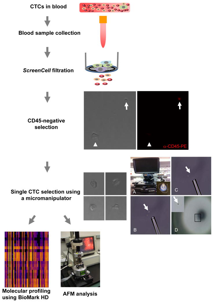
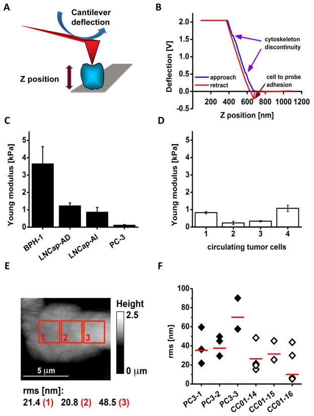
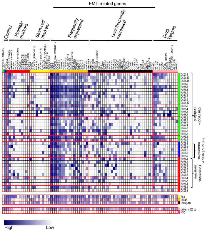
Methods: A microfiltration system was used to trap CTCs from peripheral blood based on size selection of large epithelial-like cells without CD45 leukocyte marker. These cells individually retrieved with a micromanipulator device were assessed for cell membrane physical properties using atomic force microscopy. Additionally, 38 CTCs from eight prostate cancer patients were used to determine expression profiles of 84 EMT-related and reference genes using a microfluidics-based PCR system.

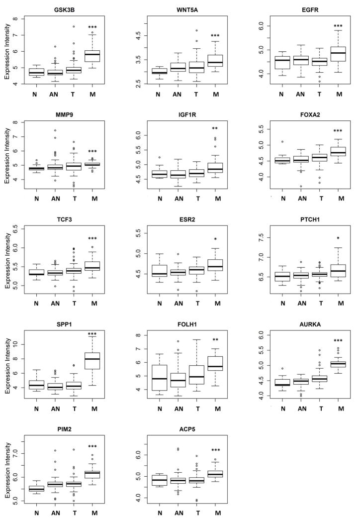
Results: Increased cell elasticity and membrane smoothness were found in CTCs compared to noncancerous cells, highlighting their potential invasiveness and mobility in the peripheral circulation. Despite heterogeneous expression patterns of individual CTCs, genes that promote mesenchymal transitioning into a more malignant state, including IGF1, IGF2, EGFR, FOXP3, and TGFB3, were commonly observed in these cells. An additional subset of EMT-related genes (e.g., PTPRN2, ALDH1, ESR2, and WNT5A) were expressed in CTCs of castration-resistant cancer, but less frequently in castration-sensitive cancer.

Conclusions: The study suggests that an incremental expression of EMT-related genes in CTCs is associated with metastatic castration-resistant cancer. Although CTCs represent a group of highly heterogeneous cells, their unique EMT-related gene signatures provide a new opportunity for personalized treatments with targeted inhibitors in advanced prostate cancer patients.


Figures

Fig. 1
Fig. 1
A schematic flow chart of CTC isolation and analyses. The details are described in the Materials and Methods section. In anti-CD45 negative selection, representative microscopic photos show a CTC (white arrow head) was negative for anti-CD45-PE staining, whereas a blood cell (white arrow) positive. In single CTC selection, the left panel shows four representative CTCs and the right panel illustrates the single cell isolation using a micromanipulator and an Evos fl microscope. A: An Evos fl microscope and a micromanipulator (inset). B: A pipette tip pointing to a cell (white arrow) selected using a micromanipulator. C: The single cell was aspirated into the pipette tip from the place it was previously located at (white arrow). D: The selected single cell was placed on a petri dish. A higher magnification of the single cell (black rectangle) is shown in the inset.
Fig. 2
Fig. 2
AFM probing of cell surface indicates that CTCs exhibit mechanical phenotype resembling highly metastatic cultured prostate cancer cells. A: A scheme illustrating the principle of measuring cell elasticity. A cell (blue) bound to a mica surface (grey) is indented by a tip (red triangle) mounted on a flexible cantilever (red board) proportionally to the cell elasticity. Deflection of the cantilever (blue arrow) changes a position of a laser beam reflection that measures force needed to indent the cell. The distance between a tip end and the cell is represented by the Z position (thick vertical arrow) directly measured by a piezoelectric element of the microscope. B: An example of a force plot of individual CTC (cell #4). Blue arrows point at positions of little humps at which the tip likely sensed a cytoskeleton discontinuity. Adhesion forces between the tip and the cell bent the cantilever in the opposite direction as indicated by the red arrow. C: Histogram comparing elasticity of four prostate cell lines. The elasticity is presented as the Young modulus. The benign BPH-1 cells are the stiffest (showed the largest Young modulus), whereas androgen dependent LNCap are more elastic followed by the LNCap androgen independent, and by PC-3 cells that are highly metastatic, and also the softest. Histograms represent mean values with corresponding SD. D: Histogram comparing elasticity of four individual CTCs. These cells were as soft as the cancerous cell lines presented in the panel C. Histograms represent mean values with corresponding SD. E: Height topography image of a single CTC (cell #4) recorded with a spherical tip in contact mode. Roughness (rms in nm) of the cell membrane calculated for three 2.5 by 2.5 μm. The cell is flat since it is tightly bound to a glass plate with poly-L-Lys and also may represent a strongly metastatic phenotype.
Fig. 3
Fig. 3
Heterogeneous expression profiles of EMT-related and other genes among CTCs. RNA from CTCs was subjected to microfluidics-based single-cell qRT-PCR analysis using a BioMark HD system. Gene expression for each gene was obtained as described in materials and methods and displayed in a blue-white gradient. Gene symbols and gene groups were labeled on the top and CTC numbers and patient groups on the right. EMT-related genes are further divided into two groups: the frequently expressed group and the less frequently expressed group.
Fig. 4
Fig. 4
Elevated cumulative expression of EMT-related genes and signaling pathways in CTCs from castration-resistant patients. Cumulative expression EMT-related genes in each CTC are displayed in box plots among CR, CR-IS and CS patients. A: Cumulative gene expression of frequently expressed EMT-related genes. B: Cumulative gene expression of less frequently expression EMT-related genes. C: Cumulative gene expressions of WNT, SHH and TGF-β signaling pathways. Data were analyzed using one-way ANOVA and unpaired Student’s t test. A p value of <0.05 is considered as statistically significant. CR: castration-resistant; CR-IS: castration-resistant and immunotherapy sensitive; CS: castration-sensitive.
Fig. 5
Fig. 5
Elevated expression of EMT-related genes and drug target genes in metastatic prostate cancer. In silico analysis of gene expression revealed that expression of nine EMT-related genes and five drug target genes are higher in clinical metastatic prostate tumors than normal prostate. Data were analyzed using Student’s t test. N: normal; AN: normal tissue adjacent to tumor; T: tumor; and M: metastatic. *, p<0.05; **, p<0.01; ***, p<0.001.

References

    1. Aguirre-Ghiso JA. Models, mechanisms and clinical evidence for cancer dormancy. Nat Rev Cancer. 2007;7:834–846. - PMC - PubMed
    1. Meng S, Tripathy D, Frenkel EP, Shete S, Naftalis EZ, Huth JF, Beitsch PD, Leitch M, Hoover S, Euhus D, Haley B, Morrison L, Fleming TP, Herlyn D, Terstappen LW, Fehm T, Tucker TF, Lane N, Wang J, Uhr JW. Circulating tumor cells in patients with breast cancer dormancy. Clin Cancer Res. 2004;10:8152–8162. - PubMed
    1. Dittmar T, Heyder C, Gloria-Maercker E, Hatzmann W, Zanker KS. Adhesion molecules and chemokines: the navigation system for circulating tumor (stem) cells to metastasize in an organ-specific manner. Clin Exp Metastasis. 2008;25:11–32. - PubMed
    1. Sun YX, Schneider A, Jung Y, Wang J, Dai J, Cook K, Osman NI, Koh-Paige AJ, Shim H, Pienta KJ, Keller ET, McCauley LK, Taichman RS. Skeletal localization and neutralization of the SDF-1(CXCL12)/CXCR4 axis blocks prostate cancer metastasis and growth in osseous sites in vivo. J Bone Miner Res. 2005;20:318–329. - PubMed
    1. Alix-Panabieres C, Schwarzenbach H, Pantel K. Circulating tumor cells and circulating tumor DNA. Annu Rev Med. 2012;63:199–215. - PubMed

Publication types

MeSH terms