. 2013 Apr;65(4):1022-31.
doi: 10.1002/art.37825.

Protein kinase Cβ is required for lupus development in Sle mice

Affiliations

Protein kinase Cβ is required for lupus development in Sle mice

David Oleksyn et al. Arthritis Rheum. 2013 Apr.

Abstract

Objective: To evaluate the requirement for protein kinase Cβ (PKCβ) in the development of lupus in mice, and to explore the potential of targeting PKCβ as a therapeutic strategy in lupus.

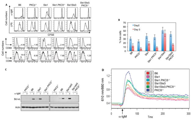
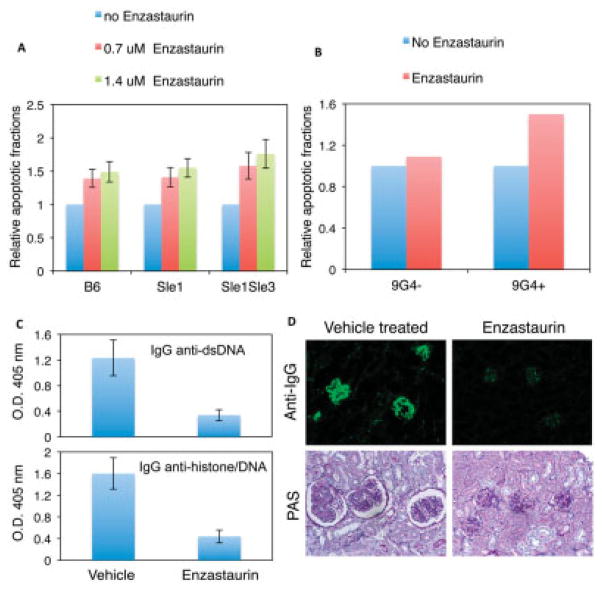
Methods: Congenic mice bearing the disease loci Sle1 or Sle1 and Sle3, which represent different stages of severity in the development of lupus, were crossed with PKCβ-deficient mice. The effect of PKCβ deficiency in lupus development was analyzed. In addition, the effects of the PKCβ-specific inhibitor enzastaurin on the survival of B cells from mice with lupus and human 9G4-positive B cells as well as the in vivo effect of enzastaurin treatment on the development of lupus in Sle mice were investigated.

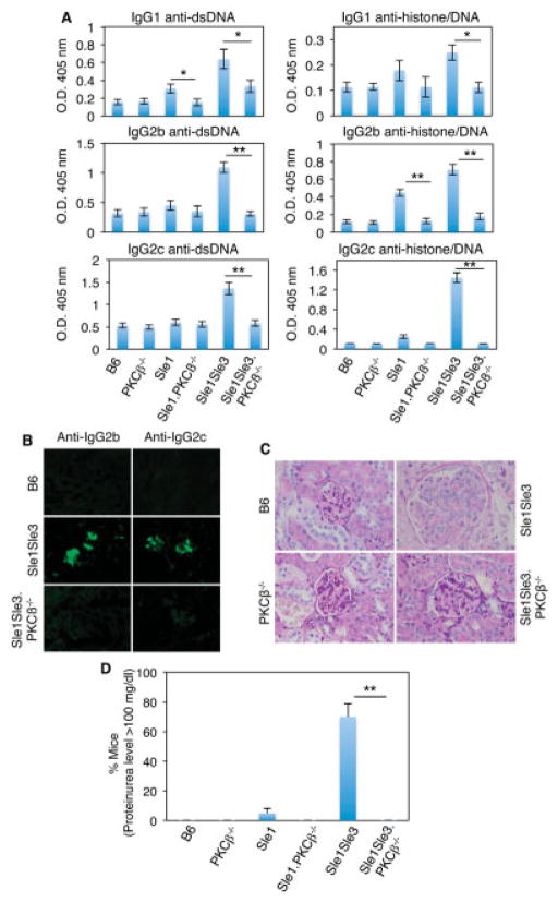
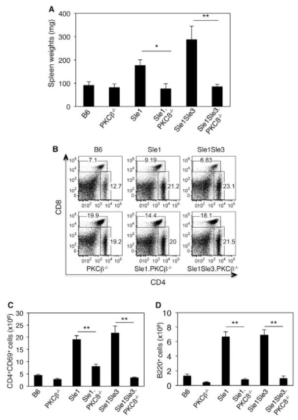
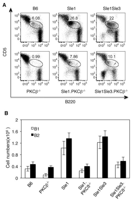
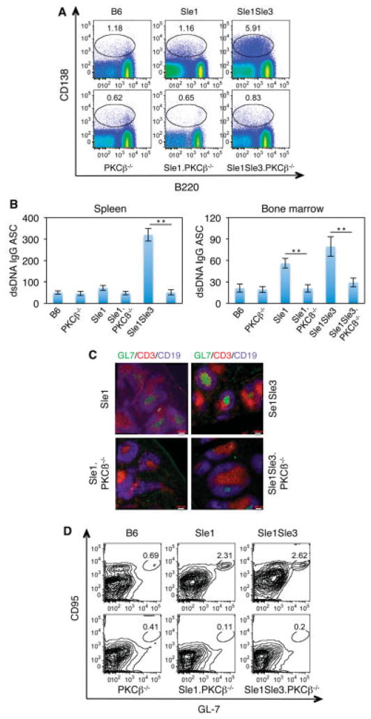
Results: In Sle mice, PKCβ deficiency abrogated lupus-associated phenotypes, including high autoantibody levels, proteinuria, and histologic features of lupus nephritis. Significant decreases in spleen size and in the peritoneal B-1 cell population, reduced numbers of activated CD4 T cells, and normalized CD4:CD8 ratios were observed. PKCβ deficiency induced an anergic B cell phenotype and preferentially inhibited autoreactive plasma cells and autoantibodies in mice with lupus. Inhibition of PKCβ enhanced apoptosis of both B cells from Sle mice and human autoreactive B cells (9G4 positive). Treatment of Sle mice with the PKCβ-specific inhibitor enzastaurin prevented the development of lupus.

Conclusion: This study identifies PKCβ as a central mediator of lupus pathogenesis, suggesting that PKCβ represents a promising therapeutic target for the treatment of systemic lupus erythematosus. Moreover, the results indicate the feasibility of using a PKCβ inhibitor for the treatment of lupus.


Figures

Figure 1
Figure 1
Protein kinase Cβ (PKCβ) deficiency prevents lupus development in Sle mice. A, Levels of serum IgG anti–double-stranded DNA (anti-dsDNA) and antihistone/anti-dsDNA autoantibodies from the indicated mouse strains (8–10 months old), as determined by enzyme-linked immunosorbent assay. For analysis of anti-dsDNA IgG and antihistone/anti-dsDNA, sera were diluted 100-fold and 200-fold, respectively. Values are the mean ± SD of ≥3 mice per group. B, Frozen kidney sections from 5-month-old mice, stained with fluorescein isothiocyanate–labeled anti-IgG2b or anti-IgG2c. C, Paraffin-embedded kidney sections from the same mice as in B, stained with periodic acid–Schiff reagent. Enlarged glomeruli that are typical of Sle1.Sle3 mice were not observed in PKCβ-deficient Sle mice. Images in B and C are representative of 2 independent experiments. D, Proteinuria levels, as measured with Uristix strips. Values are the mean ± SD of 8–10 mice per strain. * = P <0.05; ** = P < 0.01. In B and C, original magnification × 20 and × 40, respectively.
Figure 2
Figure 2
Protein kinase Cβ (PKCβ) deficiency reverses splenomegaly and restores the CD4:CD8 T cell ratio in Sle mice. A, Spleen weights in the indicated mouse strains at age 8 months. B, Representative results of flow cytometric analysis of spleen T cells from 5–6-month-old mice. Cells were stained with CD4 and CD8 antibodies. C and D, Absolute numbers of activated CD69+ cells in the CD4+ T cell population (C) and in the B220+ cell population (D). Results shown in B, C, and D were obtained from the same group of mice. Bars in A, C, and D show the mean ± SD of 5 mice per group (A) and 3 mice per group (C and D). * = P < 0.05; ** = P < 0.01.
Figure 3
Figure 3
Protein kinase Cβ (PKCβ) deficiency decreases the number of peritoneal B-1 cells in Sle mice. A, Flow cytometric analysis of peritoneal B-1 cells from the indicated mouse strains (5–6 months old). The cells were stained with B220 and CD5 antibodies. The encircled areas show B-1a cells. Results are representative of 3 independent experiments. B, Absolute numbers of B-1a (CD5+B220low) and B-2 (B220+CD23+) cells in the peritonea of the indicated mouse strains. Bars show the mean ± SD of 3 independent experiments.
Figure 4
Figure 4
PKCβ deficiency results in a reduction in plasma cell numbers and abrogates spontaneous germinal cell (GC) formation in Sle mice. A, Flow cytometric analysis of splenocytes from 5–6-month-old PKCβ-deficient Sle mice. Cells were stained with the indicated antibodies. The encircled areas show spleen plasmablasts (CD138highB220+) and plasma cells (CD138highB220low/negative). Results are representative of 3 independent experiments. B, Expression of IgG anti-dsDNA plasma cells (antibody-secreting cells [ASCs]) in 1 × 10⁶ cells from spleen or bone marrow, as determined by enzyme-linked immunospot assay. Bars show the mean ± SD. ** = P < 0.01. C, Representative immunofluorescence images of spontaneous GC (CD19+GL7+) formation in spleen sections. Spleen sections from the indicated mouse strains were stained with GL7 (green), CD3 (red), and CD19 (blue). Original magnification × 20. D, Representative results of flow cytometric analysis of GC cells (CD19+CD95+GL7+) in the indicated mouse strains. Results shown in A, B, and C were obtained from the same group of mice. See Figure 1 for other definitions.
Figure 5
Figure 5
Protein kinase Cβ (PKCβ) deficiency impairs the B cell receptor response and survival of B cells from Sle mice. A, In vitro responses of B cells to anti-IgM or lipopolysaccharide (LPS) stimulation. Splenocytes from 6-month-old mice were labeled with 5,6-carboxyfluorescein succinimidyl ester (CFSE) (shaded area) and stimulated with anti-IgM or LPS or were left in the medium without any added stimuli for 48 hours (black lines). Top, PKCβ deletion reduced the proliferative response of B cells to anti-IgM stimulation but not LPS stimulation. Bottom, PKCβ deficiency reduced up-regulation of CD69 and CD86 expression, as determined by fluorescence-activated cell sorting analysis. B, Effect of PKCβ deficiency on cell viability. Purified spleen B cells from the indicated mouse strains were incubated in RPMI 1640 medium. Cell viability was assayed by flow cytometry, using an annexin V detection kit. Bars show the mean ± SD. C, Expression of Bcl-xL in anti-IgM–stimulated splenocytes from the indicated mouse strains, as analyzed by Western blotting. Actin was used as a loading control. D, Effect of PKCβ deletion on calcium signaling in B cells from Sle mice. Splenocytes from the indicated mouse strains were loaded with 1 μM Fura Red to evaluate their ability to flux Ca2+ in response to anti-IgM. B220+ cells were gated for the analysis. All data shown are representative of at least 2 independent experiments.
Figure 6
Figure 6
The protein kinase Cβ (PKCβ)–specific inhibitor enzastaurin induces apoptosis of lupus B cells and prevents lupus development in Sle mice. A, Effect of enzastaurin on apoptosis of lupus B cells. Purified splenic B cells were treated with anti-IgM antibody in the presence or absence (control) of enzastaurin for 48 hours and analyzed with an annexin V detection kit. The fractions of annexin V–positive (apoptotic) cells in the samples treated with only anti-IgM (control) are set at 1. B, Sensitivity of human 9G4-positive and 9G4-negative B cells to PKCβ inhibition. Purified splenic B cells were treated with enzastaurin for 24 hours. The apoptotic fractions from untreated samples are set at 1. Results are representative of 2 independent experiments. C, Levels of serum IgG anti–double-stranded DNA (anti-dsDNA) and antihistone/anti-dsDNA autoantibodies from vehicle-treated control mice and enzastaurin-treated mice, as analyzed by enzyme-linked immunosorbent assay. Bars in A–C show the mean ± SD of 3 independent experiments. D, Representative immunofluorescent images of IgG deposition (top) and glomeruli (bottom) in kidney sections from Sle1.Sle3 mice treated with vehicle or enzastaurin. Original magnification × 20 (top); × 40 (bottom). PAS = periodic acid–Schiff.

References

    1. Sanz I, Lee FE. B cells as therapeutic targets in SLE. Nat Rev Rheumatol. 2010;6:326–37. - PMC - PubMed
    1. Looney RJ, Anolik J, Sanz I. A perspective on B-cell-targeting therapy for SLE. Mod Rheumatol. 2010;20:1–10. - PMC - PubMed
    1. Doreau A, Belot A, Bastid J, Riche B, Trescol-Biemont MC, Ranchin B, et al. Interleukin 17 acts in synergy with B cell-activating factor to influence B cell biology and the pathophysiology of systemic lupus erythematosus. Nat Immunol. 2009;10:778–85. - PubMed
    1. Stadanlick JE, Kaileh M, Karnell FG, Scholz JL, Miller JP, Quinn WJ, III, et al. Tonic B cell antigen receptor signals supply an NF-κB substrate for prosurvival BLyS signaling. Nat Immunol. 2008;9:1379–87. - PMC - PubMed
    1. Cancro MP. Signalling crosstalk in B cells: managing worth and need. Nat Rev Immunol. 2009;9:657–61. - PMC - PubMed

Publication types

MeSH terms