. 2012 Dec;72(6):902-11.
doi: 10.1002/ana.23689.

N-methyl-D-aspartate receptor antibodies in herpes simplex encephalitis

Affiliations

N-methyl-D-aspartate receptor antibodies in herpes simplex encephalitis

Harald Prüss et al. Ann Neurol. 2012 Dec.

Abstract

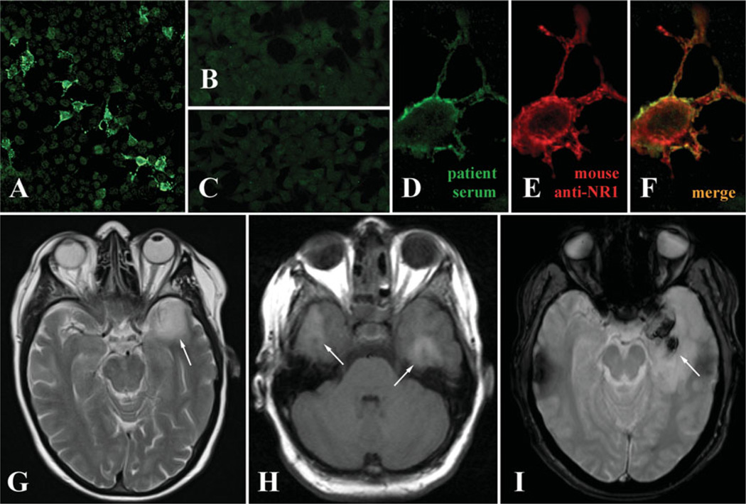
Objective: To determine the presence and kinetics of antibodies against synaptic proteins in patients with herpes simplex virus encephalitis (HSE).

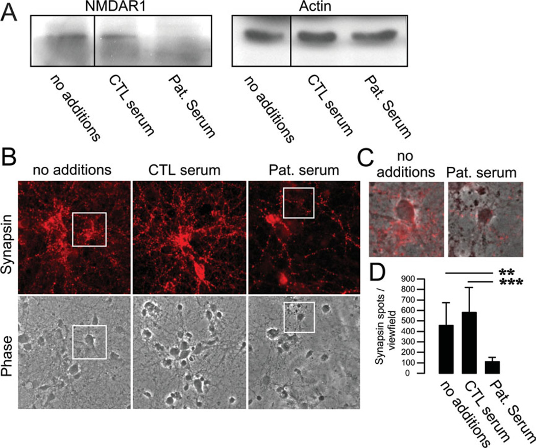
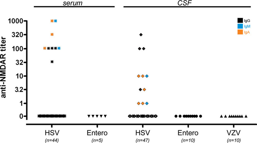
Methods: Retrospective analysis of 44 patients with polymerase chain reaction-proven HSE for the presence of a large panel of onconeuronal and synaptic receptor antibodies. The effect of patients' serum was studied in cultures of primary mouse hippocampal neurons.

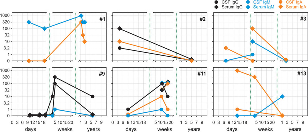
Results: N-Methyl-D-aspartate receptor (NMDAR) antibodies of the immunoglobulin (Ig) subtypes IgA, IgG, or IgM were detected in 13 of 44 patients (30%) in the course of HSE, suggesting secondary autoimmune mechanisms. NMDAR antibodies were often present at hospital admission, but in some patients developed after the first week of HSE. Antibody-positive sera resulted in downregulation of synaptic marker proteins in hippocampal neurons.

Interpretation: Some patients with HSE develop IgA, IgG, or IgM autoantibodies against NMDAR. Sera from these patients alter the density of neuronal synaptic markers, suggesting a potential pathogenic disease-modifying effect. These findings have implications for the understanding of autoimmunity in infectious diseases, and prospective studies should reveal whether the subgroup of patients with HSE and NMDAR antibodies may benefit from immunotherapy. .


Conflict of interest statement

Potential Conflicts of Interest

C.K.: employment, Euroimmun. C.P.: employment, stock/ stock options, Euroimmun. K.B.: employment, Euroimmun. L.K.: employment, Euroimmun. W.S.: board membership, employment, stock/stock options, Euroimmun. J.D.: grants/grants pending, Euroimmun, NIH/ NCI; patents, Athena Diagnostics, Euroimmun. K.-P.W.: employment, stock/stock options, Euroimmun

Figures

FIGURE 1
FIGURE 1
N-Methyl-d-aspartate receptor (NMDAR) autoantibodies in herpes simplex encephalitis (HSE) patients. (A) Immunopositive staining of transfected HEK cells overexpressing the NR1 subunit of NMDARs when probed with patient serum and anti-immunoglobulin (Ig)M secondary antibodies. (B) No staining is observed in control-transfected cells. (C) No staining is observed of transfected cells probed with IgM-positive serum, but an anti-IgG secondary antibody. (D–F) Higher magnification of NR1-transfected cells demonstrating colabeling of patient IgM and a murine anti-NR1 antibody (Biomol International, Plymouth Meeting, PA; dilution, 1:1,000). (G–I) The brain magnetic resonance imaging of patients with HSE (G) can be indistinguishable from imaging in NMDAR encephalitis (H) with predominant affection of the temporal lobes (arrows). However, large hemorrhagic changes in the temporal lobes are typical for HSE (I). [Color figure can be viewed in the online issue, which is available at annalsofneurology.org.]
FIGURE 2
FIGURE 2
Downregulation of membrane N-methyl-d-aspartate receptors (NMDARs) and synaptic proteins by patient serum containing NMDAR immunoglobulin (Ig)M antibodies. (A) Primary mouse hippocampal neurons were incubated for 3 days with patient serum (1:100 dilution), and the membrane fraction was run in Western blots. Staining against NR1 subunits revealed a strong downregulation of NMDARs following incubation with IgM-positive patient serum (Pat. serum). Incubation with control serum (CTL serum) or media (no additions) had no effect on NMDAR expression. Actin was used for loading control. (B) Immunostaining of hippocampal neurons incubated with patient serum for 3 days led to a dramatic reduction of synapsin-positive clusters. (C) Higher magnification and overlay of the insets in B. (D) Quantification of synapsin spots demonstrating profound downregulation after treatment with NMDAR antibody-positive serum. ** p < 0.005, *** p < 0.001. [Color figure can be viewed in the online issue, which is available at annalsofneurology.org.]
FIGURE 3
FIGURE 3
Differential kinetics of N-methyl-d-aspartate receptor (NMDAR) antibody titers of immunoglobulin (Ig)A, IgG, or IgM subtypes in herpes simplex encephalitis patients (see also Table 1). The development of NMDAR antibody titers during the disease course followed a heterogeneous pattern (day 0 = hospital admission). In most cases, NMDAR antibodies were present already within the first days of initial clinical presentation (eg, patient 2), too early for a primary immune response and indicative of pre-existing antibodies. However, in some cases antibodies did not evolve before the first week (eg, patient 9), suggesting a newly stimulated B cell-mediated response. Late follow-up (after up to 7 years) generally demonstrated reduction in antibody titers in serum and cerebrospinal fluid (CSF; eg, patients 2, 3, 9, 13). [Color figure can be viewed in the online issue, which is available at annalsofneurology.org.]
FIGURE 4
FIGURE 4
N-Methyl-d-aspartate receptor (NMDAR) antibodies in encephalitis patients. Of patients with herpes simplex virus (HSV) encephalitis, 20.5% had immunoglobulin (Ig)G, IgM, or IgA NMDAR antibodies in serum and 23.4% in cerebrospinal fluid (CSF), whereas no antibodies were detected in the CSF and serum of patients with enterovirus encephalitis or CSF of varicella zoster virus (VZV) encephalitis (serum of VZV cases not available). [Color figure can be viewed in the online issue, which is available at annalsofneurology.org.]

References

    1. Steiner I, Kennedy PG, Pachner AR. The neurotropic herpes viruses: herpes simplex and varicella-zoster. Lancet Neurol. 2007;6:1015–1028. - PubMed
    1. Whitley R. Herpes simplex virus. In: Sheld W, Whitley R, C Marra, editors. Infections of the central nervous system. Philadelphia, PA: Lippincott Williams & Wilkins; 2004. pp. 123–144.
    1. Raschilas F, Wolff M, Delatour F, et al. Outcome of and prognostic factors for herpes simplex encephalitis in adult patients: results of a multicenter study. Clin Infect Dis. 2002;35:254–260. - PubMed
    1. Sellner J, Dvorak F, Zhou Y, et al. Acute and long-term alteration of chemokine mRNA expression after anti-viral and anti-inflammatory treatment in herpes simplex virus encephalitis. Neurosci Lett. 2005;374:197–202. - PubMed
    1. Kamei S, Sekizawa T, Shiota H, et al. Evaluation of combination therapy using aciclovir and corticosteroid in adult patients with herpes simplex virus encephalitis. J Neurol Neurosurg Psychiatry. 2005;76:1544–1549. - PMC - PubMed

Publication types

MeSH terms