. 2013 Feb;73(2):246-58.
doi: 10.1002/ana.23780. Epub 2012 Dec 31.

Early interneuron dysfunction in ALS: insights from a mutant sod1 zebrafish model

Affiliations
Free PMC article

Early interneuron dysfunction in ALS: insights from a mutant sod1 zebrafish model

Alexander McGown et al. Ann Neurol. 2013 Feb.
Free PMC article

Abstract

Objective: To determine, when, how, and which neurons initiate the onset of pathophysiology in amyotrophic lateral sclerosis (ALS) using a transgenic mutant sod1 zebrafish model and identify neuroprotective drugs.

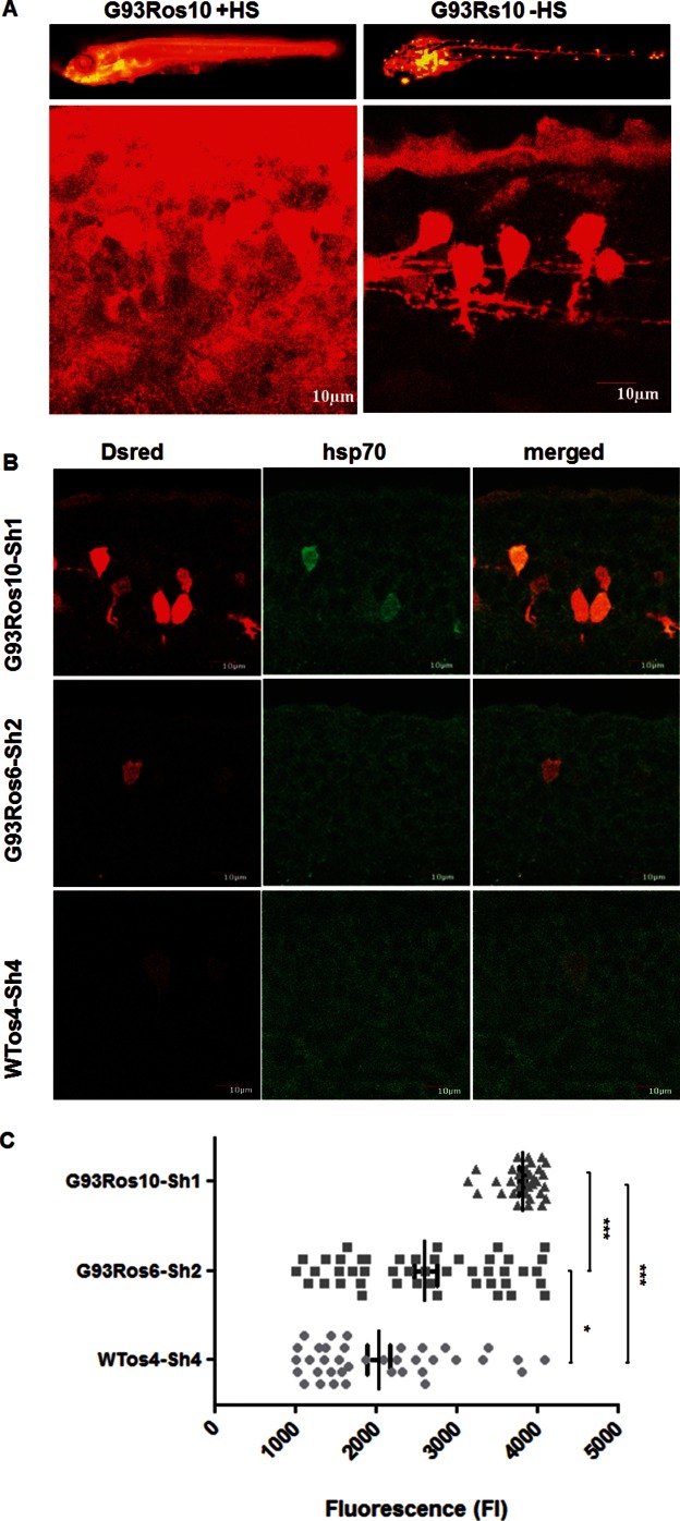
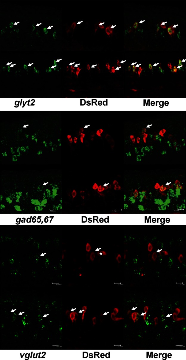
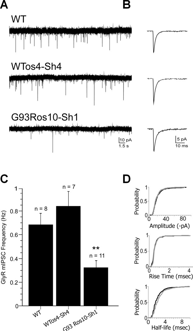
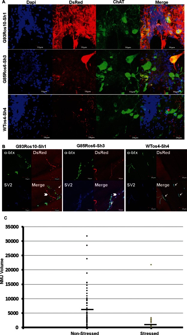
Methods: Proteinopathies such as ALS involve mutant proteins that misfold and activate the heat shock stress response (HSR). The HSR is indicative of neuronal stress, and we used a fluorescent hsp70-DsRed reporter in our transgenic zebrafish to track neuronal stress and to measure functional changes in neurons and muscle over the course of the disease.

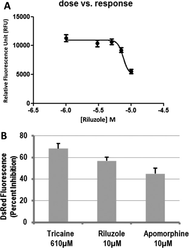
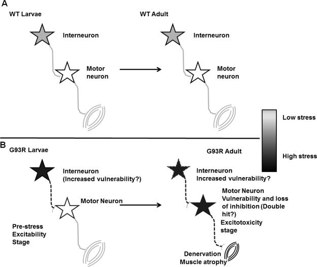
Results: We show that mutant sod1 fish first exhibited the HSR in glycinergic interneurons at 24 hours postfertilization (hpf). By 96 hpf, we observed a significant reduction in spontaneous glycinergic currents induced in spinal motor neurons. The loss of inhibition was followed by increased stress in the motor neurons of symptomatic adults and concurrent morphological changes at the neuromuscular junction (NMJ) indicative of denervation. Riluzole, the only approved ALS drug and apomorphine, an NRF2 activator, reduced the observed early neuronal stress response.

Interpretation: The earliest event in the pathophysiology of ALS in the mutant sod1 zebrafish model involves neuronal stress in inhibitory interneurons, resulting from mutant Sod1 expression. This is followed by a reduction in inhibitory input to motor neurons. The loss of inhibitory input may contribute to the later development of neuronal stress in motor neurons and concurrent inability to maintain the NMJ. Riluzole, the approved drug for use in ALS, modulates neuronal stress in interneurons, indicating a novel mechanism of riluzole action.


Figures

FIGURE 1
FIGURE 1
Sod1 transgenic fish induce the heat shock response (HSR) without exposure to heat stress. (A) Live G93Ros10-Sh1 showing induction of hsp70 measured by DsRed fluorescence at 7 days postfertilization in whole embryos (top panel) and 30 hours postfertilization (hpf) spinal cord (bottom panel). Larvae were heat shocked (+HS) or left unexposed to heat shock (−HS), and images of HSR induction were compared. When exposed to heat, the larvae showed global induction of the HSR (left panel), whereas in the absence of heat shock, only neuronal, neuroepithelial, and occasionally muscle cells show induction of the HSR (right panel). (B) Multiple mutant lines G93Ros10-Sh1 and G93Ros6-Sh2 show HSR induction in the absence of heat shock. Confocal images of spinal neurons show induction of endogenous hsp70 (middle column) in high expresser G93Ros10 line (top panel) in the same cells that showed strong DsRed expression (top row, left column). Moderate expresser G93Ros6-Sh2 line (middle row) and high expresser WTos4-Sh4 line (bottom row) do not show elevated hsp70 levels above background. (C) Quantitation of the DsRed fluorescence in individual neurons in the zebrafish embryonic spinal cord (30 hpf) by average fluorescence intensity. Average fluorescence of individual DsRed-positive neurons was measured and analyzed by analysis of variance. *p < 0.05 for G93Ros6-Sh2 and WTos4-Sh4; ***p < 0.0001 for G93Ros10-Sh1 and WTos4-Sh4. Size bars = 10μM. [Color figure can be viewed in the online issue, which is available at http://www.annalsofneurology.org.]
FIGURE 2
FIGURE 2
Embryonic mutant sod1 zebrafish show induction of neuronal stress predominantly in the spinal inhibitory glycinergic interneurons. In situ hybridization with probes for inhibitory and excitatory neurons (left panels) and DsRed riboprobes (middle panel). Top panel: glyt2-positive glycinergic inhibitory interneurons; middle panel: gad65,67-positive γ-aminobutyric acidergic inhibitory interneurons (dorsal longitudinal ascending and some glycinergic interneurons); bottom panel: vglut2-positive excitatory interneurons (commissural primary ascending and commissural secondary ascending interneurons) that cross-modulate the spinal locomotor circuitry. Time: 24 hours postfertilization. Size bars = 10μM. [Color figure can be viewed in theonline issue, which is available at http://www.annalsofneurology.org.]
FIGURE 3
FIGURE 3
Reduced glycinergic transmission onto motor neurons of sod1 zebrafish larvae. (A) Representative traces depicting voltage clamp (holding potential = −75mV) recordings of spontaneous glycinergic miniature postsynaptic currents (mPSCs) in motor neurons of wild-type (WT), WT Sod1 overexpresser (WTos4-Sh4), and sod1 mutant (G93Ros10-Sh1) fish at 4 days postfertilization. Downward deflections represent occasional quantal release of glycine from presynaptic terminals. (B) Average of 30 consecutive glycinergic mPSCs from each experimental condition. (C) Bar chart depicting mean mPSC frequency for each experimental condition. GlyR = glycine receptor; mIPSC = miniature inhibitory postsynaptic current. **WT vs G93R p < 0.001. (D) Cumulative probability plots of mPSC amplitude, rise time, and half-life (p < 0.05) in WT (black lines) and G93Ros10-Sh1 (gray lines) motor neurons.
FIGURE 4
FIGURE 4
Mutant sod1 zebrafish show stress in the large spinal motor neurons of the adult spinal cord and concurrent loss of neuromuscular junction (NMJ). (A) Spinal cord cross sections from 1- to 1.5-year-old adult zebrafish stained with 4′,6-diamidino-2-phenylindole (Dapi), DsRed antibody, and ChAT antibody show robust induction of the heat shock stress response in spinal cord motor neurons. DsRed colocalized with ChAT in the high expresser (×3) G93Ros10-Sh1 line (top panel) and the moderate expresser (×2) G85Ros6-Sh3 line (middle panel). The high expresser (×3) WTos4-Sh4 line shows little DsRed expression, and DsRed did not colocalize with the large ChAT-positive motor neurons (bottom panel). (B) Muscle sections labeled with synaptic vesicle-2 (SV2) antibody (blue), α-bungarotoxin(α-btx) (green), and DsRed (red) in high expresser G93Ros10-Sh1 (left panel), low expresser G85Ros6-Sh3 (middle panel), and high expresser WTos4-Sh4 (right panel). Normal NMJs are indicated by arrows. Abnormal NMJs (arrowheads) where pre- and postsynaptic markers are absent or small and punctate were detected in the muscle sections from the mutant lines (left and middle panels) but not in the high expresser wild-type line (right panel). (C) One hundred thirteen NMJs from multiple sections were measured for NMJ volume from confocal stacks across multiple planes in SV2-positive–DsRed-negative axons and SV2-positive–DsRed-positive axons using colocalization software from National Institutes of Health Image J and analyzed by unpaired t test. Significant reduction in NMJ volume was observed associated with stressed motor axons as compared to the nonstressed axons. The mean is represented as a line over the distribution. Each dot represents the volume of an individual NMJ. p < 0.00001. [Color figure can be viewed in the online issue, which is available at http://www.annalsofneurology.org.]
FIGURE 5
FIGURE 5
Inhibition of the stress response in sod1 G93Ros10-Sh1 zebrafish embryos by riluzole and NRF2 activator R-apomorphine. (A) Dose–response curve showing dose-dependent inhibition of the stress response by riluzole in sod1 G93Ros10-Sh1 embryos treated for 4 days with 1, 3, 5, 7, and 10μM riluzole, p < 0.001. (B) Percentage inhibition of the stress response expressed by reduction in DsRed fluorescence in embryos treated with 610μM Tricaine (p < 0.000001), 10μM riluzole (p < 0.00001), and 10μM R-apomorphine (p < 0.001) as compared to 0.1% dimethyl sulfoxide–treated embryos. Mean ± standard error of the mean.
FIGURE 6
FIGURE 6
Model of neuronal stress propagation in zebrafish model of amyotrophic lateral sclerosis. (A) Although wild-type (WT) Sod1 overexpresser zebrafish show some interneuron stress in early development, the stress levels are low and there is no propagation of the stress response to motor neurons. (B) Mutant sod1 transgenic zebrafish show stress initially in the inhibitory interneurons that cause dysfunction of glycinergic inhibitory interneurons with reduced glycinergic input to motor neurons. This lack of inhibition may be a factor contributing to the stress response developing in motor neurons in the adult zebrafish. Stressed motor neurons are unable to maintain synaptic function with resulting synaptic withdrawal at the neuromuscular junction (NMJ). The retraction of the presynaptic input at the NMJ may lead to a loss of trophic support from muscle, resulting in a vicious cycle of injury to the motor neuron, eventually leading to motor neuron degeneration and muscle atrophy. Shades of gray indicate stress levels.

Similar articles

Cited by

References

    1. Jucker M, Walker LC. Pathogenic protein seeding in Alzheimer disease and other neurodegenerative disorders. Ann Neurol. 2011;70:532–540. - PMC - PubMed
    1. Turner BJ, Talbot K. Transgenics, toxicity and therapeutics in rodent models of mutant SOD1-mediated familial ALS. Prog Neurobiol. 2008;85:94–134. - PubMed
    1. Bosco DA, Morfini G, Karabacak NM, et al. Wild-type and mutant SOD1 share an aberrant conformation and a common pathogenic pathway in ALS. Nat Neurosci. 2010;13:1396–1403. - PMC - PubMed
    1. Guareschi S, Cova E, Cereda C, et al. An over-oxidized form of superoxide dismutase found in sporadic amyotrophic lateral sclerosis with bulbar onset shares a toxic mechanism with mutant SOD1. Proc Natl Acad Sci U S A. 2012;109:5074–5079. - PMC - PubMed
    1. Polymenidou M, Cleveland DW. The seeds of neurodegeneration: prion-like spreading in ALS. Cell. 2011;147:498–508. - PMC - PubMed

Publication types

MeSH terms