. 2013 Mar;22(3):296-305.
doi: 10.1002/pro.2211. Epub 2013 Jan 17.

Insulin analog with additional disulfide bond has increased stability and preserved activity

Affiliations

Insulin analog with additional disulfide bond has increased stability and preserved activity

Tine N Vinther et al. Protein Sci. 2013 Mar.

Abstract

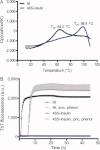
Insulin is a key hormone controlling glucose homeostasis. All known vertebrate insulin analogs have a classical structure with three 100% conserved disulfide bonds that are essential for structural stability and thus the function of insulin. It might be hypothesized that an additional disulfide bond may enhance insulin structural stability which would be highly desirable in a pharmaceutical use. To address this hypothesis, we designed insulin with an additional interchain disulfide bond in positions A10/B4 based on Cα-Cα distances, solvent exposure, and side-chain orientation in human insulin (HI) structure. This insulin analog had increased affinity for the insulin receptor and apparently augmented glucodynamic potency in a normal rat model compared with HI. Addition of the disulfide bond also resulted in a 34.6°C increase in melting temperature and prevented insulin fibril formation under high physical stress even though the C-terminus of the B-chain thought to be directly involved in fibril formation was not modified. Importantly, this analog was capable of forming hexamer upon Zn addition as typical for wild-type insulin and its crystal structure showed only minor deviations from the classical insulin structure. Furthermore, the additional disulfide bond prevented this insulin analog from adopting the R-state conformation and thus showing that the R-state conformation is not a prerequisite for binding to insulin receptor as previously suggested. In summary, this is the first example of an insulin analog featuring a fourth disulfide bond with increased structural stability and retained function.


Figures

Figure 1
Figure 1
Crystal structure of the 4SS-insulin analog. (A) Overlay of the crystal structure of the 4SS-insulin analog (blue) and HI (grey) (pdb code 1MSO.PDB), in a hexamer assembly. (B) The structure of the 4SS-insulin analog (pdb code 4EFX) with the A-chain in grey and B-chain in blue. The additional disulphide bond between B4C and A10C is shown by yellow stick representation. The 2fo-Fc electron density from the additional disulphide bond is rendered in blue at 1.0σ level. The N- and C-terminus of the B-chain are labeled. (C) Ribbon representation of the 4SS-insulin analog (blue) compared with 17 insulin structure obtained from the Protein Data Bank codes 1IZB, 1M5A, 1MSO, 1OS3, 2A3G, 2INS, 2ZP6, 3EXX, 3FHP, 3FQ9, 3ILG, 3INC, 3INS, 3RTO, 3TT8, 4E7T, and 4INS. The chains are depicted separately for clarity. The two B-chains are shown in the middle, flanked by their respective A-chain. (D) The hydrogen bonding between the A (grey) and B (blue) chains formed by introduction of the additional disulphide bond. (E) Comparison of the N-terminal part of the B-chain with that of HI (1MSO.PDB). The positions of the residues in the 4SS-insulin analog are shifted one position relative to the HI. Labels refer to sequence numbering of the HI/4SS-insulin structures, respectively. (F) The orientation of the HisB5 side chain in the 4SS-insulin analog deviates from the 17 structures compared. The HisB5 in the 4SS-insulin analog point away from the N-terminal α-helix and is not active in the helical capping which may exist in the other insulin structures. The additional disulphide bond between CysB4 and CysA10 is shown by yellow stick representation.
Figure 2
Figure 2
Probing phenol induced changes of the conformational state of insulin in the presence of zinc and sodium chloride. Phenol titration of HI with zinc (•), HI with zinc and sodium chloride (○), 4SS-insulin with zinc (▪), and 4SS-insulin with zinc and sodium chloride (□). The molar elipticity is indicative of the conformational state such that an amplitude at −2.0 M−1 cm−1 and −8.0M−1 cm−1 is indicative of T-state and R-state, respectively.
Figure 3
Figure 3
Representative insulin receptor binding curves. Representative curves for HI (•) and the 4SS-insulin (▪) with number of assays, n = 3. Each point on the graph represents the mean ± SD, n = 4 within one assay.
Figure 4
Figure 4
In vivo activity of the 4SS-insulin analog. (A) Blood glucose profiles following i.v. administration of HI and 4SS-insulin. The two analogs were administered in two doses in anaesthetized wistar rats; 1.2 nmol/kg HI (○), 3.6 nmol/kg HI (•), 1.2 nmol/kg 4SS-insulin (□), 3.6 nmol/kg 4SS-insulin (▪), and vehicle (▾) mean values ± SEM, n = 6, 5, 6, 6, 6. (B) Blood glucose lowering effect (area over curve (AOC) with the background subtracted) after i.v. administration of 4SS-insulin, HI or vehicle in fed anaesthetized wistar rats. The P value from a student two-tailed t-test between HI and the 4SS-insulin is indicated for each dose.
Figure 5
Figure 5
Stability of the 4SS-insulin analog. DSC thermographs for HI (black) and the 4SS-insulin analog (grey) recorded from 10−110°C with an increase of 1°C/min (B) ThT assay for HI (black), HI with 3Zn/hexamer and 30 mM phenol (gray), the 4SS-insulin analog (dark blue), and the 4SS-insulin analog with 3Zn/hexamer and 30 mM phenol (light blue). SD is indicated for each measure point, n = 4. The analog was subjected to continuously shaking for 45 h at 37°C.

References

    1. World Health Organization. Definition, Diagnosis and Classification of Diabetes Mellitus and its Complications (1999) Part 1: Diagnosis and Classification of Diabetes Mellitus. WHO Consultation. Geneva: World Health Organization, Department of Noncommunicable Disease Surveillance;
    1. Baker EN, Blundell TL, Cutfield JF, Cutfield SM, Dodson EJ, Dodson GG, Hodgkin DM, Hubbard RE, Isaacs NW, Reynolds CD, Sakabe K, Sakabe N, Vijayan NM. The structure of 2Zn pig insulin crystals at 1.5 A resolution. Philos Trans R Soc Lond B Biol Sci. 1988;319:369–456. - PubMed
    1. Blundell T, Dodson G, Hodgkin D, Mercola D. Insulin: The structure in the crystal and its reflection in chemistry and biology. Adv Protein Chem. 1972;26:279–402.
    1. Nicol DS, Smith LF. Amino-acid sequence of human insulin. Nature. 1960;187:483–485. - PubMed
    1. Ryle AP, Sanger F, Smith LF, Kitai R. The disulphide bonds of insulin. Biochem J. 1955;60:541–556. - PMC - PubMed

Publication types

MeSH terms

Associated data