

Spatial variability and changes of metabolite concentrations in the cortico-spinal tract in multiple sclerosis using coronal CSI
- PMID: 23281189
- PMCID: PMC4238834
- DOI: 10.1002/hbm.22229
Spatial variability and changes of metabolite concentrations in the cortico-spinal tract in multiple sclerosis using coronal CSI
Abstract
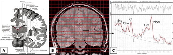
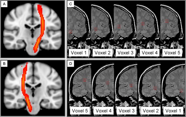
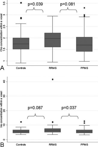
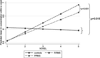
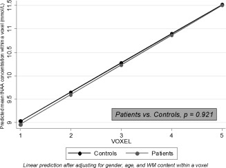
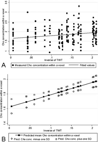
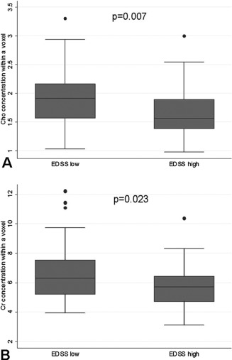
We characterized metabolic changes along the cortico-spinal tract (CST) in multiple sclerosis (MS) patients using a novel application of chemical shift imaging (CSI) and considering the spatial variation of metabolite levels. Thirteen relapsing-remitting (RR) and 13 primary-progressive (PP) MS patients and 16 controls underwent (1)H-MR CSI, which was applied to coronal-oblique scans to sample the entire CST. The concentrations of the main metabolites, i.e., N-acetyl-aspartate, myo-Inositol (Ins), choline containing compounds (Cho) and creatine and phosphocreatine (Cr), were calculated within voxels placed in regions where the CST is located, from cerebral peduncle to corona radiata. Differences in metabolite concentrations between groups and associations between metabolite concentrations and disability were investigated, allowing for the spatial variability of metabolite concentrations in the statistical model. RRMS patients showed higher CST Cho concentration than controls, and higher CST Ins concentration than PPMS, suggesting greater inflammation and glial proliferation in the RR than in the PP course. In RRMS, a significant, albeit modest, association between greater Ins concentration and greater disability suggested that gliosis may be relevant to disability. In PPMS, lower CST Cho and Cr concentrations correlated with greater disability, suggesting that in the progressive stage of the disease, inflammation declines and energy metabolism reduces. Attention to the spatial variation of metabolite concentrations made it possible to detect in patients a greater increase in Cr concentration towards the superior voxels as compared to controls and a stronger association between Cho and disability, suggesting that this step improves our ability to identify clinically relevant metabolic changes.
Keywords: MRI; MRS; multiple sclerosis.
Copyright © 2012 Wiley Periodicals, Inc.
Figures
References
-
- Armitage P, Berry G, Matthews J (2002): Statistical methods in medical research, 4th ed. Blackwell Science Ltd.
-
- Barker PB, Bonekamp D, Riedy G, Smith M (2006): Quantitation of NAA in the brain by magnetic resonance spectroscopy. Adv Exp Med Biol 576:183–197. - PubMed
-
- Bramow S, Frischer JM, Lassmann H, Koch‐Henriksen N, Lucchinetti CF, Sorensen PS, Laursen H (2010): Demyelination versus remyelination in progressive multiple sclerosis. Brain 133:2983–2998. - PubMed
Publication types
MeSH terms
Substances
Grants and funding
LinkOut - more resources
Full Text Sources
Medical
Miscellaneous
