. 2013 Feb;70(2):101-120.
doi: 10.1002/cm.21094. Epub 2012 Dec 26.

Conserved structural motifs in the central pair complex of eukaryotic flagella

Affiliations

Conserved structural motifs in the central pair complex of eukaryotic flagella

Blanca I Carbajal-González et al. Cytoskeleton (Hoboken). 2013 Feb.

Abstract

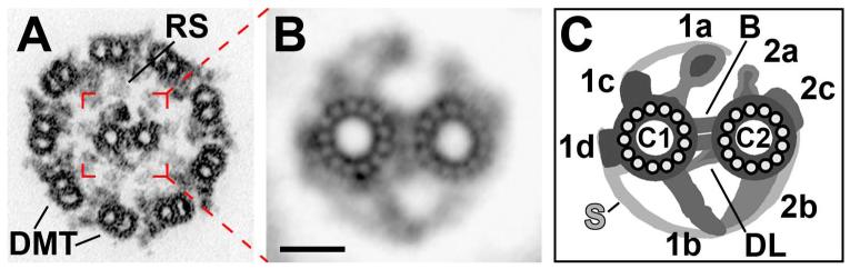
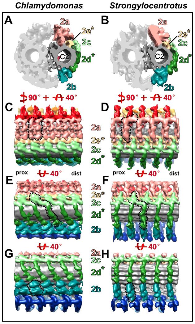
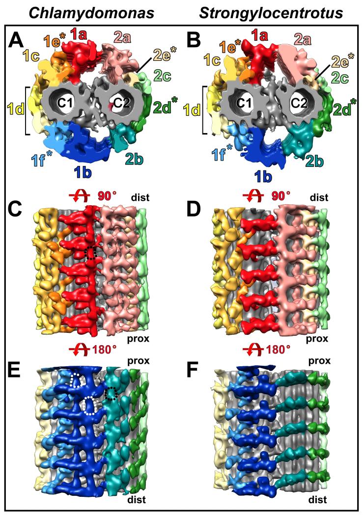
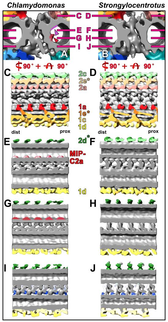
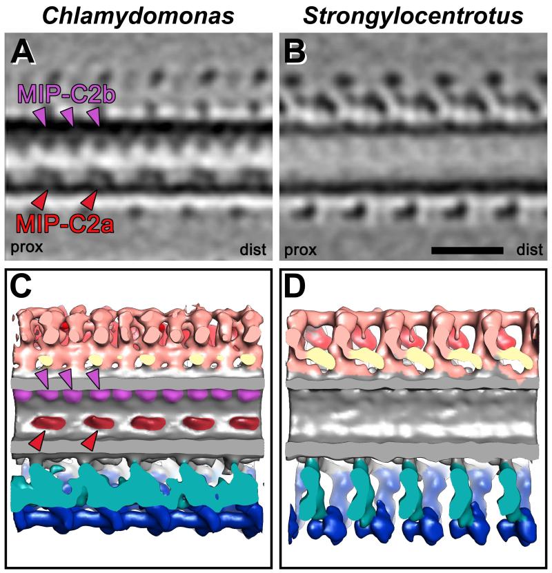
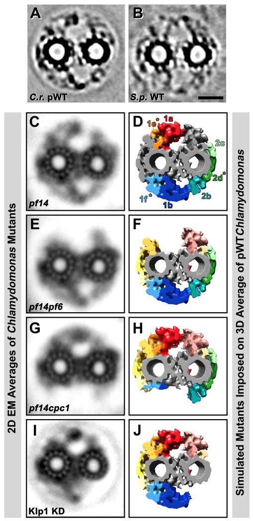
Cilia and flagella are conserved hair-like appendages of eukaryotic cells that function as sensing and motility generating organelles. Motility is driven by thousands of axonemal dyneins that require precise regulation. One essential motility regulator is the central pair complex (CPC) and many CPC defects cause paralysis of cilia/flagella. Several human diseases, such as immotile cilia syndrome, show CPC abnormalities, but little is known about the detailed three-dimensional (3D) structure and function of the CPC. The CPC is located in the center of typical [9+2] cilia/flagella and is composed of two singlet microtubules (MTs), each with a set of associated projections that extend toward the surrounding nine doublet MTs. Using cryo-electron tomography coupled with subtomogram averaging, we visualized and compared the 3D structures of the CPC in both the green alga Chlamydomonas and the sea urchin Strongylocentrotus at the highest resolution published to date. Despite the evolutionary distance between these species, their CPCs exhibit remarkable structural conservation. We identified several new projections, including those that form the elusive sheath, and show that the bridge has a more complex architecture than previously thought. Organism-specific differences include the presence of MT inner proteins in Chlamydomonas, but not Strongylocentrotus, and different overall outlines of the highly connected projection network, which forms a round-shaped cylinder in algae, but is more oval in sea urchin. These differences could be adaptations to the mechanical requirements of the rotating CPC in Chlamydomonas, compared to the Strongylocentrotus CPC which has a fixed orientation.


Figures

Fig. 1
Fig. 1. Organization of the CPC
(A-C) A cross-section of a Chlamydomonas axoneme (A) shows that the CPC is located in the center of the axoneme surrounded by the nine doublet MTs (DMT). The CPC is composed of two singlet MTs, C1 and C2, and its associated projections which extend toward the radial spokes (RS). Projection designations were determined previously using 2D EM averages (B) and are summarized in a simplified schematic model (C). Scale bar in (B) is 20 nm. All images were adapted from [Mitchell and Sale, 1999] with minor modifications.
Fig. 2
Fig. 2. Comparison of the C1 MT projections between Chlamydomonas and Strongylocentrotus
(A-H) Isosurface renderings show the C1 MT projections from CPC averages in cross-sectional (A and B) and longitudinal views (C-H), and demonstrate that the C1 projections of Chlamydomonas (A, C, E, and G) bear strong resemblance to those of Strongylocentrotus (B, D, F, and H). Only a few structural differences between Chlamydomonas and Strongylocentrotus C1 projections are present as indicated by dotted outlines in (C, D and G). Note, that the most distal portions of the 1e projection appear to be oriented in opposite directions (compare C and D), and the 1a and 1b projections are longitudinally connected in Chlamydomonas (C and G). The 1a and 1b projections repeat every 16 nm, the remaining 1d, 1c, 1e and 1f projections repeat every 32 nm giving the CPC an overall periodicity of 32 nm. Note the arches (black arrowhead in G) in the 1f projection every 32 nm. An ~80 nm long piece of the CPC (~ 2.5 repeat units) is displayed to highlight the repetitive organization of the CPC. Note that the Chlamydomonas CPC isosurface rendering was obtained from two separate averages of the C1 and C2 MT (see Supporting Information Fig. S1 and Materials and Methods section for details). This averaging approach, color coding of projections and the shown views are preserved in all following Figures. Asterisks highlight projection designations that are described in this study for the first time. For orientation, the proximal (prox) and distal (dist) side of the averages are indicated in (C and D).
Fig. 3
Fig. 3. Comparison of the C2 MT projections between Chlamydomonas and Strongylocentrotus
(A-H) Cross-sectional (A and B) and longitudinal views (C-H) of isosurface renderings from averaged CPCs show overall a high similarity in the C2 MT projections between Chlamydomonas and Strongylocentrotus, but also reveal differences outlined by black dotted lines in (C-H). In both Chlamydomonas (A, C, E, and G) and Strongylocentrotus (B, D, F, and H) all C2 projections repeat every 16 nm. Projection 2a appears more intricate in Chlamydomonas (C) than in Strongylocentrotus (D), and exhibits a “pseudo-8 nm periodicity” in the alga (C). Note that the 2d projection is angled distally in Chlamydomonas (outlined in E), but runs nearly perpendicular to the MT in Strongylocentrotus (outlined in F). The Chlamydomonas 2b projection (outlined in G) appears broader than in Strongylocentrotus (outlined in H). Projections described in this study for the first time are highlighted by asterisks. The proximal (prox) and distal (dist) side of the averages are indicated in (C and D) for easier orientation.
Fig. 4
Fig. 4. Connections between C1 and C2 projections
(A-F) Isosurface renderings from Chlamydomonas and Strongylocentrotus CPC averages show cross-sectional (A and B) and longitudinal views from the top (C and D) and bottom (E and F) of the CPC to highlight connections between C1 and C2 projections. In both organisms the C1 and C2 MTs are linked directly via a complex network of connections, called the bridge (see Fig. 5). Additional connections between the C1 and C2 halves of the CPC are formed by links between the 1a and 2a projections (C and D), as well as between the 1b and 2b projections (E and F). In Chlamydomonas, the 1a projections are also interconnected longitudinally along the MT axis (black outline in C), while they are not connected in Strongylocentrotus (D). Similarly, longitudinal connections between adjacent 1b and 2b projections are present in Chlamydomonas (black and white dotted outlines in E), but not in Strongylocentrotus (F). The proximal (prox) and distal (dist) side of the CPC are indicated for better orientation in (C-F).
Fig. 5
Fig. 5. Comparison of the C1-C2 bridge structures between Chlamydomonas and Strongylocentrotus
(A-J) Isosurface renderings from CPC averages of Chlamydomonas (A, C, E, G, and I) and Strongylocentrotus (B, D, F, H, and J) display the bridge structure in cross-sectional (A, B) and four different longitudinal views observed from the top (C and D), middle (E-H), and bottom (I and J). A complex network with several distinct connections links the C1 and C2 MTs. It is apparent that some bridge components in Chlamydomonas are arranged diagonally (I) to the MT whereas they are arranged perpendicularly to the MT in Strongylocentrotus (J). All bridge structures appear to have a 16 nm periodicity. Note the positions of the two MT inner proteins MIP-C2a and MIP-C2b (red and purple arrowheads in A) that are only present in Chlamydomonas (A, E and G; see also Fig. 6), but absent from Strongylocentrotus (B, F and H). Magenta lines in (A and B) indicate the orientation of the longitudinal views shown in (C-J). Asterisks highlight newly designated projections and the proximal (prox) and distal (dist) side is indicated to facilitate orientation in (C and D).
Fig. 6
Fig. 6. MT Inner Proteins in the C2 MT of the Chlamydomonas CPC
(A-D) Tomographic slices (A and B) and isosurface renderings (C and D) of the Chlamydomonas (A and C) and Strongylocentrotus CPC averages (B and D) reveal the presence of two MT Inner Proteins, MIP-C2a and MIP-C2b, inside the C2 MT of Chlamydomonas (red and purple arrowheads in A and C), but not in Strongylocentrotus (B and D). The larger MIP-C2a (red arrowheads) shows a 16 nm periodicity, whereas the smaller MIP-C2b (purple arrowheads) repeats every 8 nm. For orientation, proximal (prox) and distal (dist) sides of the averages are indicated in (A and B). Scale bar (B): 25 nm.
Fig. 7
Fig. 7. Classical 2D EM averages of CPC mutants and simulation of corresponding 3D structural defects
(A-J) Cross-sectional projection images through ~80 nm thick tomographic slices of CPC averages from Chlamydomonas pseudo-wildtype (C.r. pWT, A) and Strongylocentrotus wildtype (S.p. WT, B) are shown for comparison with 2D EM averages of ~80 nm thick plastic sections of the CPC from four Chlamydomonas mutants: pf14 (C) is a mutant lacking the radial spokes, but without CPC defects, i.e. the 2D averages looks like wildtype (compare with Fig. 1B). Note the similarities in densities amongst (A and C), as well as (B). The pf14pf6 (E), pf14cpc1 (G) double mutants and the Klp1 knockdown (I) all show CPC defects. Isosurface renderings of the Chlamydomonas pWT average have been edited by deleting selected projection densities (D, F, H and J) to reflect densities likely to be absent in the depicted Chlamydomonas CPC mutants (C, E, G, and I). The pf14pf6 double mutant in (E) appears to lack projections 1a and 1e. The 1b and 1f projections are almost completely absent in the pf14cpc1 double mutant (G and H). From the Kinesin-like protein 1 knockdown (Klp1 KD) (I) the 2b-d projections appear to be absent (J), which includes Hydin (see Fig. 8). Scale bar (B): 25 nm. Images from (C, E, and G) were adapted from [Mitchell and Sale, 1999] and image (I) was modified from [Yokoyama et al., 2004].
Fig. 8
Fig. 8. Biochemical analysis of Chlamydomonas mutations affecting the 1b and 2b projections
Axonemes from wildtype (WT), cpc1-1 mutant and Klp1-RNAi knockdown (AC92) strains were blotted with different antibodies (left labels) to reveal the level of specific central pair proteins. Hydin is somewhat reduced in axonemes of the cpc1-1 mutant, as previously described [Lechtreck and Witman, 2007], and completely missing from axonemes from Klp1 knockdown strain AC92. The effect of Klp1 knockdown on Hydin has not been reported previously although it is consistent with the loss of the 2b projection in this strain [Yokoyama et. al., 2004]. Klp1 is unaffected in axonemes from the cpc1-1 mutant, and reduced in the knockdown strain. Outer row dynein intermediate chain IC2 was used as a loading control.

References

    1. Adams GMW, Huang B, Piperno G, Luck DJL. Central-pair microtubular complex of Chlamydomonas flagella: Polypeptide composition as revealed by analysis of mutants. J Cell Biol. 1981;91:69–76. - PMC - PubMed
    1. Afzelius BA. Cilia-related diseases. J Path. 2004;204:470–477. - PMC - PubMed
    1. Barber CF, Heuser T, Carbajal-González BI, Botchkarev VV, Jr., Nicastro D. Three-dimensional structure of the radial spokes reveals heterogeneity and interactions with dyneins in Chlamydomonas flagella. Mol Biol Cell. 2012;23:111–120. - PMC - PubMed
    1. Bernstein M, Beech PL, Katz SG, Rosenbaum JL. A new kinesin-like protein (Klp1) localized to a single microtubule of the Chlamydomonas flagellum. J Cell Biol. 1994;125(6):1313–1326. - PMC - PubMed
    1. Branche C, Kohl L, Toutirais G, Buisson J, Cosson J, Bastin P. Conserved and specific functions of axoneme components in trypanosome motility. J Cell Sci. 2006;119(Pt 16):3443–3455. - PubMed

Publication types

MeSH terms

LinkOut - more resources