. 2013 Feb;123(2):580-93.
doi: 10.1172/JCI65013. Epub 2013 Jan 2.

An obligate cell-intrinsic function for CD28 in Tregs

Affiliations

An obligate cell-intrinsic function for CD28 in Tregs

Ruan Zhang et al. J Clin Invest. 2013 Feb.

Abstract

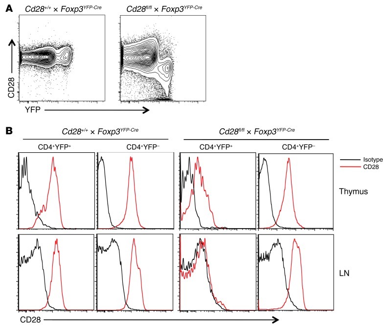
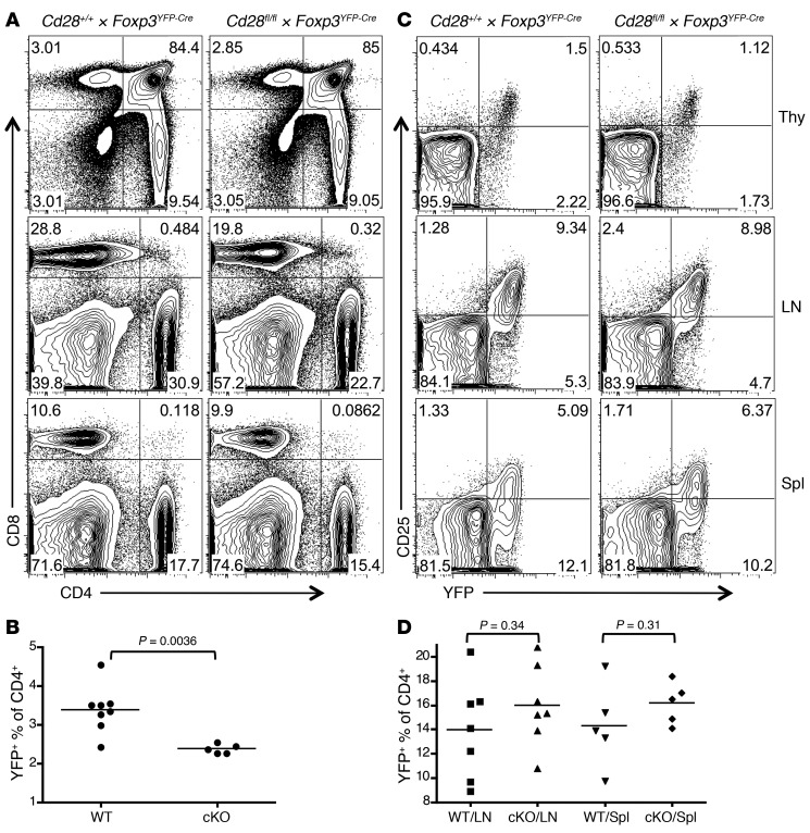
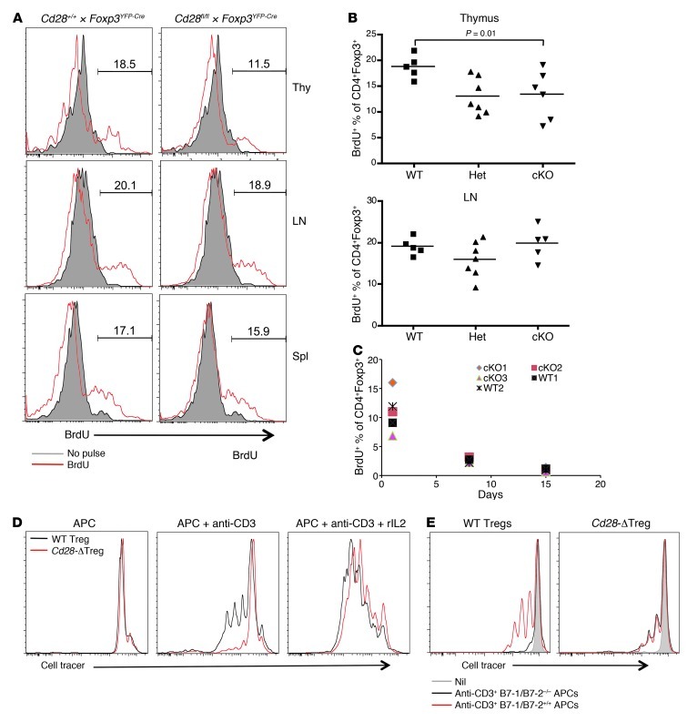
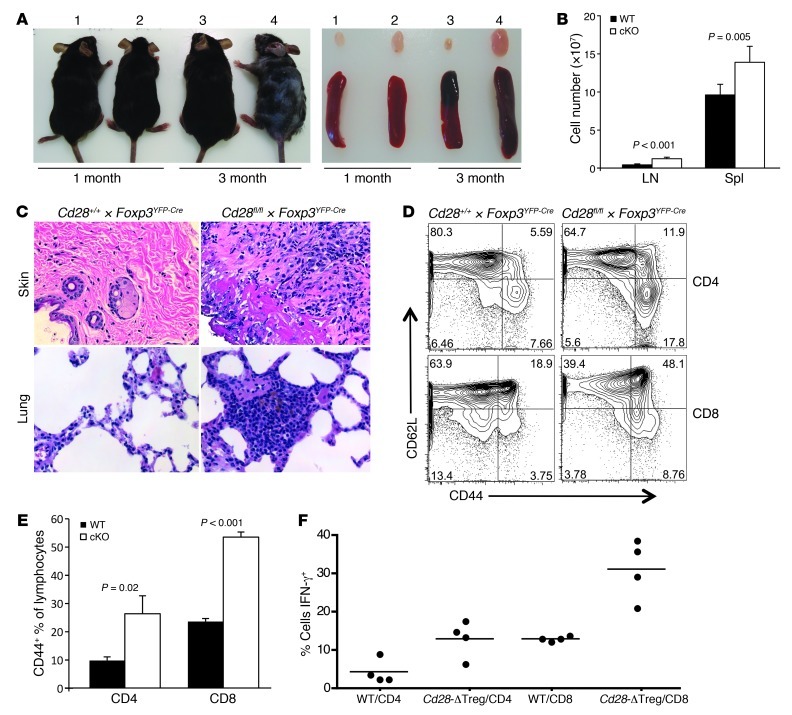
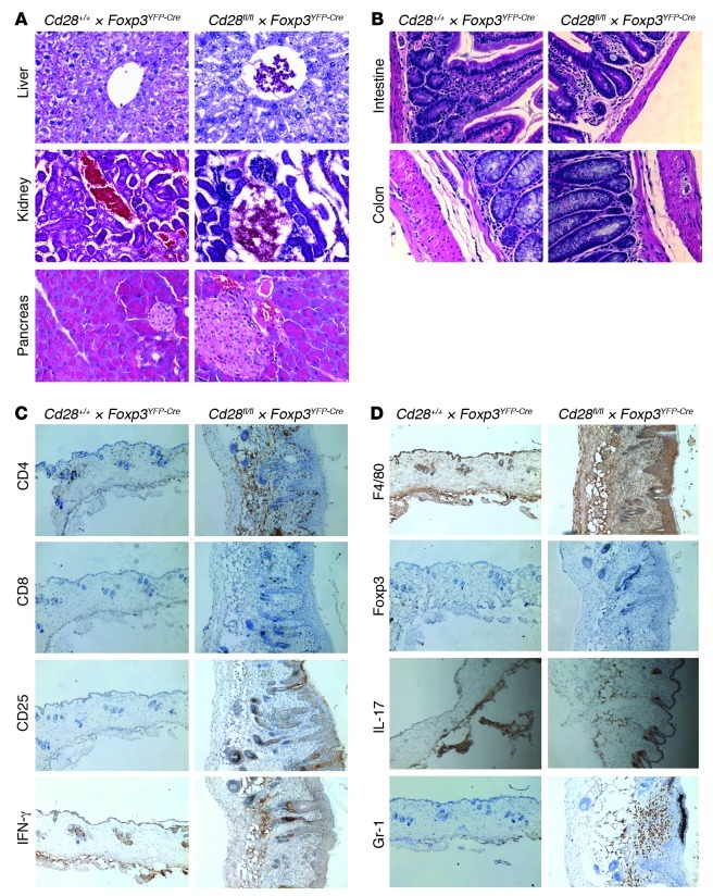
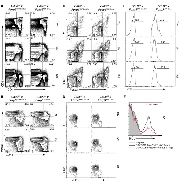
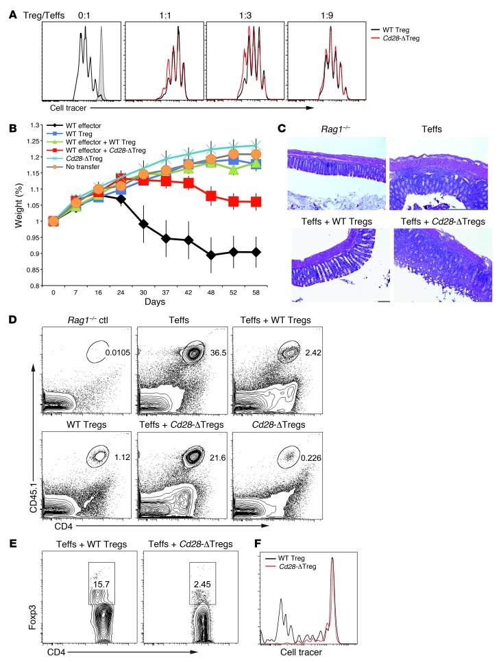
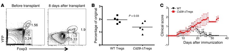
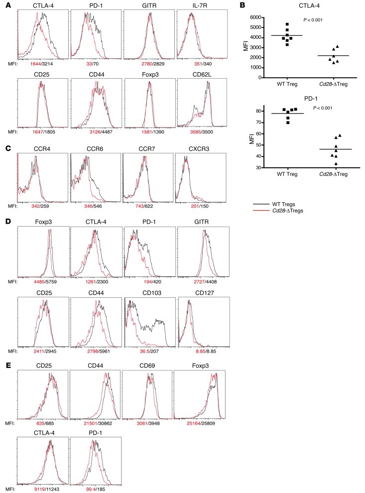
Tregs expressing the transcription factor FOXP3 are critical for immune homeostasis. The costimulatory molecule CD28 is required for optimal activation and function of naive T cells; however, its role in Treg function has been difficult to dissect, as CD28 is required for thymic Treg development, and blockade of CD28-ligand interactions has confounding effects in trans on nonregulatory cells. To address this question, we created Treg-specific Cd28 conditional knockout mice. Despite the presence of normal numbers of FOXP3+ cells, these animals accumulated large numbers of activated T cells, developed severe autoimmunity that primarily affected the skin and lungs, and failed to appropriately resolve induced experimental allergic encephalomyelitis. This in vivo functional impairment was accompanied by dampened expression of CTLA-4, PD-1, and CCR6. Disease occurrence was not due to subversion of Cd28-deficient Tregs into pathogenic cells, as complementation with normal Tregs prevented disease occurrence. Interestingly, in these "competitive" environments, Cd28-deficient Tregs exhibited a pronounced proliferative/survival disadvantage. These data demonstrate clear postmaturational roles for CD28 in FOXP3+ Tregs and provide mechanisms which we believe to be novel to explain how interruption of CD28-ligand interactions may enhance immune responses independent of effects on thymic development or on other cell types.


Figures

Figure 1
Figure 1. CD28 expression in control and Cd28-ΔTreg mice.
(A) CD28 and YFP expression in gated CD4+ lymph node cells. (B) CD28 expression in the indicated cells from thymus or lymph node. Analyses in A and B are from 6-week-old littermates and are representative of 3 independent experiments and litters.
Figure 2
Figure 2. Treg development and homeostasis in Cd28-ΔTreg mice.
(A and B) Representative analysis for (A) CD4 and CD8 and (B) CD25 and YFP in the thymi (Thy), lymph nodes, and spleens (Spl) of littermate control (Cd28+/+ × Foxp3YFP-Cre) and Cd28-ΔTreg mice (Cd28fl/fl × Foxp3YFP-Cre) mice (age 5 weeks). (C and D) Percentage of Tregs (defined as YFP+) in CD4+CD8 T cells from (C) thymi and (D) lymph nodes or spleens of 4- to 7-week-old mice. WT, control Cd28+/+ × Foxp3YFP-Cre mice; cKO, Cd28fl/fl × Foxp3YFP-Cre mice. Symbols represent individual mice; horizontal bars indicate the mean.
Figure 3
Figure 3. Treg homeostasis.
(A and B) BrdU incorporation assay. 4- to 7-week-old WT or Cd28-ΔTreg mice were pulsed with BrdU every 12 hours for 3 days and sacrificed, and relevant tissues were analyzed by flow cytometry. Representative BrdU staining of gated CD4+FOXP3+ cells is shown in A, and results of each experiment is plotted in B (Het, male Cd28fl/+ × Foxp3YFP-Cre mice; cKO, male Cd28fl/fl × Foxp3YFP-Cre mice). Symbols represent individual mice; horizontal bars indicate the mean. (C) BrdU incorporation in thymectomized mice, as per the protocol in A. CD4+FOXP3+ cells from blood were analyzed at the indicated times after the last BrdU pulse. (D and E) Sorted WT Tregs or Cd28-ΔTregs were labeled with CellTrace Violet and were stimulated by soluble CD3 plus mitomycin-treated T cell–depleted splenocytes (D) with or without rIL-2 or (E) with B7-1/B7-2 double-knockout T cell–depleted splenocytes.
Figure 4
Figure 4. Cd28-ΔTreg mice develop autoimmunity with skin inflammation.
(A) Gross appearance and lymph nodes and spleens from representative WT littermate control (labeled 1 and 3) and Cd28-ΔTreg (labeled 2 and 4) mice. (B) Cell numbers in lymph nodes and spleens from 2- to 5-month-old mice. 6 littermate pairs were analyzed. Data represent mean ± SEM. (C) H&E staining of skin and lung tissue in a 3-month-old Cd28-ΔTreg mouse. Original magnification, ×400. (D) CD44+CD62Lhi cells in lymph nodes of a representative 4-month-old Cd28-ΔTreg mice. (E) Proportions of CD44+ cells from lymph nodes of 2-month-old mice. 5 littermate pairs were analyzed. Data represent mean ± SEM. (F) Percentage of IFN-γ+ cells in stimulated splenocytes from 2- to 5-month-old littermate pairs. Symbols represent individual mice; horizontal bars indicate the mean.
Figure 5
Figure 5. Histologic analysis.
(A and B) H&E staining of (A) livers, kidneys, and pancreata or (B) intestines and colons in 3-month-old Cd28-ΔTreg mice. (C and D) Immunohistochemistry staining of skin tissues from a 3-month-old Cd28-ΔTreg mice. Original magnification, ×400 (A and B); ×100 (C and D). Data are representative of at least 3 animals.
Figure 6
Figure 6. Analysis of female mice heterozygous for Foxp3YFP-Cre.
(AD) Flow cytometric analyses of thymus, lymph node, and spleen of a representative 3-month-old Cd28fl/flFoxp3YFP-Cre/+ female mouse and female littermate control. CD4+ cells are gated in B (similar findings were observed in a CD8+ gate; data not shown) and C, and CD4+FOXP3+CD25+ cells are gated in D. (E) Percentage of YFP+ Cd28-ΔTregs in a representative Cd28fl/flFoxp3YFP-Cre/+ female mouse and a Cd28fl/+Foxp3YFP-Cre/YFP-Cre female littermate control. In the former animal, depending on which X chromosome is randomly inactivated, some cells will delete CD28, and others will not. In these mice, YFP+ Tregs are Cd28–/–, while YFP Tregs are Cd28+/+. In the control, as the animals are heterozygous for the targeted CD28 allele but homozygous for Foxp3YFP-Cre, all Tregs in these mice will be Cd28+/–. In both cases, CD4+CD25+FOXP3+ lymph node cells are shown. (F) Cd28fl/flFoxp3YFP-Cre/+ female mice were administered BrdU as in Figure 3, and BrdU staining of YFP+ and YPF cells was separately analyzed.
Figure 7
Figure 7. Cd28-ΔTreg development in bone marrow chimeras.
B6 mice (CD45.2+Thy1.1+) were irradiated and reconstituted with a 1:1 ratio of bone marrow cells from WT B6 (CD45.1+Thy1.2+) and Cd28-ΔTreg mice (CD45.2+Thy1.2+). Blood cells were analyzed 6 months later, and CD4+ cells were gated. Note that the WT mice did not carry the Foxp3YFP-Cre transgene. Thus, Tregs in the WT mice are CD25+YFP, while in the Cd28-ΔTreg mice, they are CD25+YFP+. A total of 7 mice were analyzed with similar results.
Figure 8
Figure 8. Suppressive function of Cd28-ΔTregs.
(A) In vitro suppression assay. Sorted WT naive cells were stimulated by soluble CD3 and T cell–depleted splenocytes with the addition of different ratios of WT Tregs or Cd28-ΔTregs. Tregs were sorted from 4-week-old mice. Teff, effector T cells. (B) In vivo colitis induction. Weight loss of Rag1–/– mice adoptively transferred with sorted CD4+CD45RBhi effector T cells with or without WT Tregs or Cd28-ΔTregs. A total of 6 mice were analyzed for each group. Data represent mean ± SEM. (C) H&E staining of the colons of mice in B. Original magnification, ×100. (D and E) Representative analysis of donor CD45.1+CD4+ effector cells and regulatory cells in the colons of mice in B. (F) Sorted WT Tregs or Cd28-ΔTregs were labeled with CellTrace Violet and adoptively transferred to Rag1–/– hosts. Lymph node cells were analyzed 7 days after transfer. Data are representative of 3 experiments.
Figure 9
Figure 9. Cd28-ΔTregs have defective accumulation and function in vivo.
(A and B) Cd28fl/flFoxp3Cre female mice were transplanted with BALB/c body skin. WT and Cd28-ΔTregs from the blood were analyzed before or 8 days after transplantation. The percentage of increase of Tregs was analyzed in B, and each dot represents 1 mouse (horizontal bars indicate the mean). (C) Clinical scores of rMOG/CFA plus pertussis toxin–induced EAE in female WT Cd28+/+ × FOXP3YFP-Cre/YFP-Cre (WT) or Cd28fl/fl × FOXP3YFP-Cre/YFP-Cre (Cd28-ΔTreg) mice. A total of 7 mice in each group are shown. Data represent mean ± SEM.
Figure 10
Figure 10. Cell surface markers in Cd28-ΔTregs.
(AC) Analysis of CD4+CD25+YFP+ cells from lymph nodes of 4-week-old Cd28fl/flFoxp3YFP-Cre male mice. Symbols represent individual mice; horizontal bars indicate the mean. (D) Comparison of WT and Cd28-ΔTregs in a representative female Cd28fl/flFoxp3YFP-Cre/+ mouse. Over 3 mice were analyzed. (E) Activation markers in sorted WT Tregs or Cd28-ΔTregs stimulated for 3 days with soluble CD3 plus mitomycin-treated T cell–depleted splenocytes. Experiments were repeated 3 times.

References

    1. Khattri R, Cox T, Yasayko SA, Ramsdell F. An essential role for Scurfin in CD4(+)CD25(+) T regulatory cells. Nat Immunol. 2003;4(4):337–342. doi: 10.1038/ni909. - DOI - PubMed
    1. Fontenot JD, Gavin MA, Rudensky AY. Foxp3 programs the development and function of CD4(+)CD25(+) regulatory T cells. Nat Immunol. 2003;4(4):330–336. doi: 10.1038/ni904. - DOI - PubMed
    1. Hori S, Nomura T, Sakaguchi S. Control of regulatory T cell development by the transcription factor Foxp3. Science. 2003;299(5609):1057–1061. doi: 10.1126/science.1079490. - DOI - PubMed
    1. Wildin RS, Smyk-Pearson S, Filipovich AH. Clinical and molecular features of the immunodysregulation, polyendocrinopathy, enteropathy, X linked (IPEX) syndrome. J Med Genet. 2002;39(8):537–545. doi: 10.1136/jmg.39.8.537. - DOI - PMC - PubMed
    1. Godfrey VL, Wilkinson JE, Russell LB. X-linked lymphoreticular disease in the scurfy (sf) mutant mouse. Am J Pathol. 1991;138(6):1379–1387. - PMC - PubMed

Publication types

MeSH terms