. 2013 Feb;123(2):712-26.
doi: 10.1172/JCI65728. Epub 2013 Jan 2.

CXCR5⁺ T helper cells mediate protective immunity against tuberculosis

Affiliations

CXCR5⁺ T helper cells mediate protective immunity against tuberculosis

Samantha R Slight et al. J Clin Invest. 2013 Feb.

Abstract

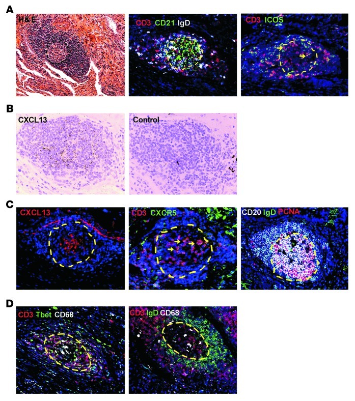
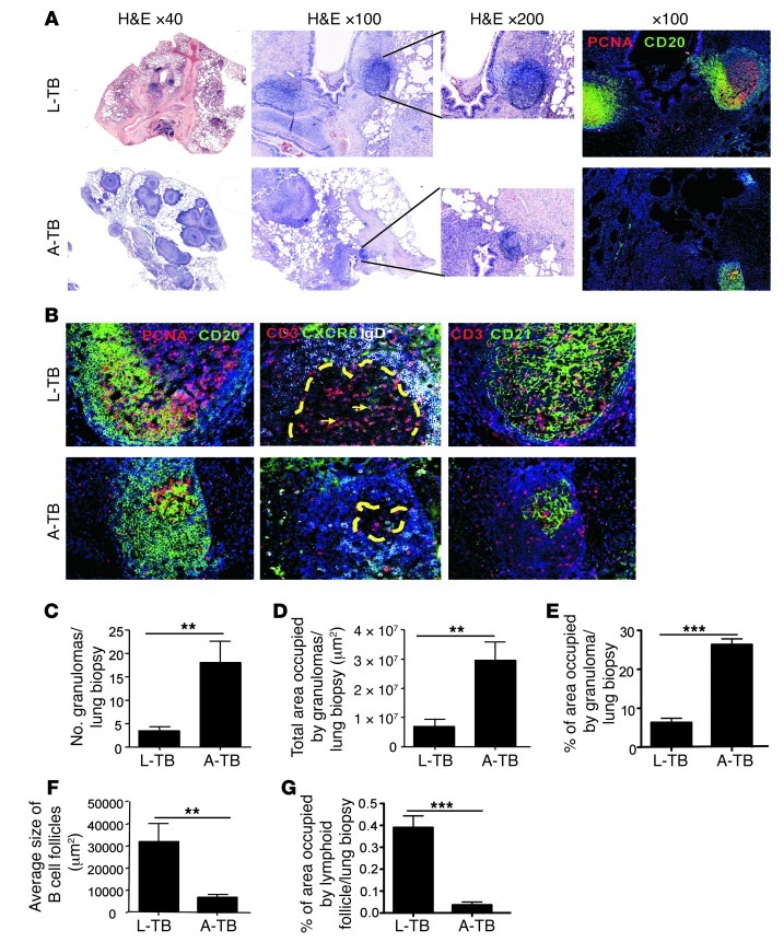
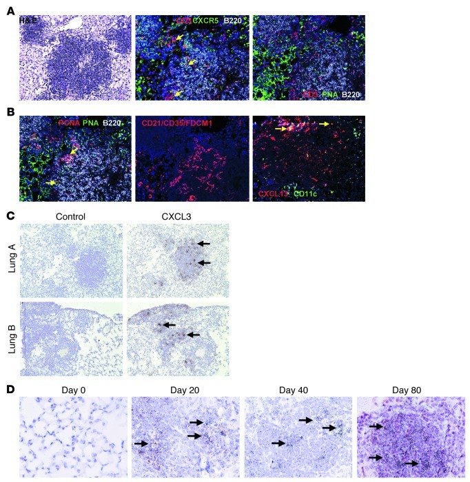
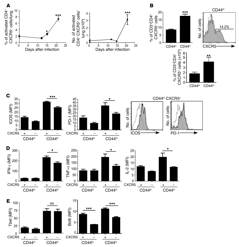
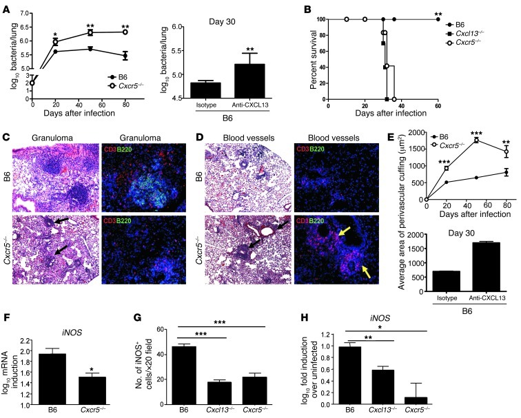
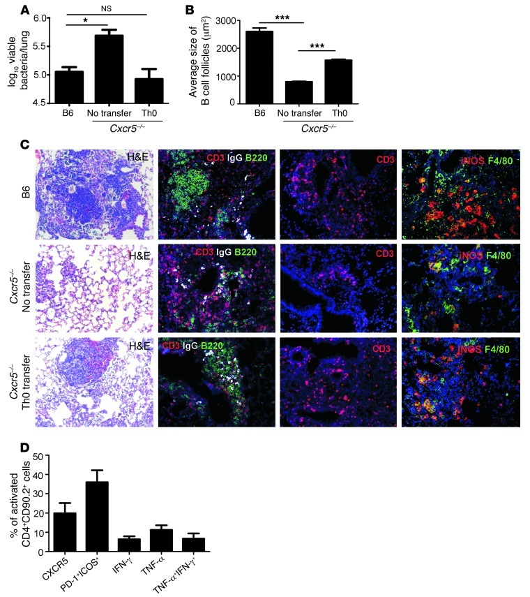
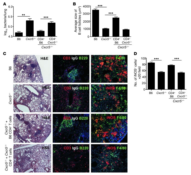
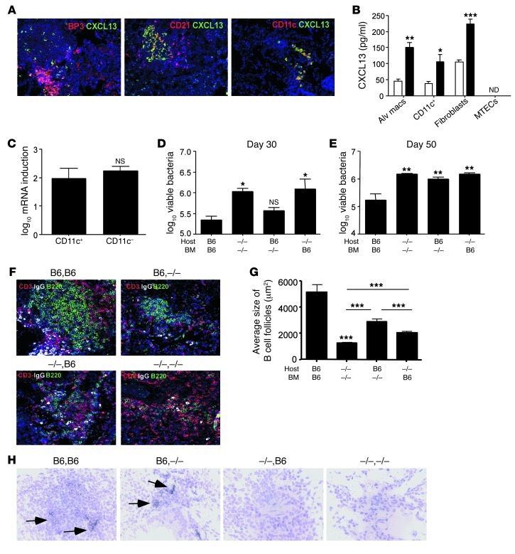
One third of the world's population is infected with Mycobacterium tuberculosis (Mtb). Although most infected people remain asymptomatic, they have a 10% lifetime risk of developing active tuberculosis (TB). Thus, the current challenge is to identify immune parameters that distinguish individuals with latent TB from those with active TB. Using human and experimental models of Mtb infection, we demonstrated that organized ectopic lymphoid structures containing CXCR5+ T cells were present in Mtb-infected lungs. In addition, we found that in experimental Mtb infection models, the presence of CXCR5+ T cells within ectopic lymphoid structures was associated with immune control. Furthermore, in a mouse model of Mtb infection, we showed that activated CD4+CXCR5+ T cells accumulated in Mtb-infected lungs and produced proinflammatory cytokines. Mice deficient in Cxcr5 had increased susceptibility to TB due to defective T cell localization within the lung parenchyma. We demonstrated that CXCR5 expression in T cells mediated correct T cell localization within TB granulomas, promoted efficient macrophage activation, protected against Mtb infection, and facilitated lymphoid follicle formation. These data demonstrate that CD4+CXCR5+ T cells play a protective role in the immune response against TB and highlight their potential use for future TB vaccine design and therapy.


Figures

Figure 1
Figure 1. CXCR5+ T cells accumulate within ectopic lymphoid structures of human TB granulomas.
Serial sections of formalin-fixed, paraffin-embedded (FFPE) lung biopsies from A-TB patients underwent H&E staining (A, left panel). CXCL13 mRNA was detected by ISH with a CXCL13 cRNA probe (B). Sections were analyzed by immunofluorescence using antibodies specific to CD3, CD21, IgD; and CD3, ICOS (A), or CXCL13; CD3, CXCR5; and PCNA, IgD, CD20 (C), or CD3, Tbet, CD68; and CD3, IgD, CD68 (D). All sections were counterstained with DAPI (blue). GC with a central core of PCNA+CD20+IgD+ large B blasts are outlined with yellow dashed lines. Yellow arrows point to typical CD3+ ICOS+ or CXCR5+ T cells (A and C). CXCL13 protein localizes inside a GC outlined with yellow dashed line (C). Original magnification, ×200; ×600 (ISH).
Figure 2
Figure 2. Ectopic lymphoid structures are associated with immune control during TB in the NHP model of Mtb infection.
NHPs aerosol infected with Mtb CDC1551 exhibited either L-TB or A-TB as described in Methods. Thirteen weeks after infection or at necropsy, lung FFPE serial sections were stained with H&E (A) or analyzed by immunofluorescence for PCNA, CD20 (A and B), or CD3, CXCR5, IgD; CD3, CD21 (B). All sections were counterstained with DAPI (blue). GC, containing large, proliferating B blasts (PCNA+CD20+), is outlined in dashed yellow lines. Yellow arrows point to typical CD3+CXCR5+ T cells within lymphoid structures. The images shown are from a typical representative section. Number of granulomas (C), total area occupied by granulomas (D), percentage of granuloma area occupied per biopsy (E), and average size and percentage of area occupied by B cell lymphoid follicles (F and G) were determined with the morphometric tool of the Zeiss Axioplan microscope. Original magnification, ×200, unless otherwise indicated. The data points represent the mean (±SD) of values from 6–15 NHPs (AG). **P = 0.005, ***P = 0.0005.
Figure 3
Figure 3. CXCR5+ T cells localize within ectopic lymphoid follicles in murine TB granulomas.
B6 mice were aerosol infected with approximately 100 CFU Mtb H37Rv, and on day 50 after infection, FFPE serial sections were stained with H&E or analyzed by immunofluorescence for CD3, CXCR5, B220; and CD3, PNA, B220 (A); PCNA, PNA, B220; CD21-CD35-FDCM1; and CD11c, CXCL13 (B). All sections were counterstained with DAPI (blue). Yellow arrows point to CD3+CXCR5+ T cells (A), PCNA+B220+ B cells (B), or CXCL13+CD11c+ cells (B). Lung sections were analyzed by ISH to determine localization of Cxcl13 mRNA expression using a murine CXCL13 cRNA probe (C) during different points of infection (D). Black arrows point to localization of Cxcl13 mRNA within granulomas. Original magnification, ×200 (A and B), ×100 (C), and ×600 (D). One of 2 or more experiments is shown.
Figure 4
Figure 4. Activated CD4+CXCR5+ T cells accumulate in the lung during Mtb infection and express both Tfh-like and Th1-like cell markers.
B6 mice were infected as in Figure 3. (A) The frequency and number of activated CD4+CXCR5+ T cells were determined by flow cytometry at different time points after infection. (B) Frequency and number of activated (CD44hi) and unactivated (CD44lo) CD4+CXCR5+ T cells were determined in Mtb-infected lungs on day 25 after infection by flow cytometry. A typical histogram showing CXCR5-specific staining (filled) within activated CD4+ T cells and relevant isotype control antibody (open) is shown (B). Expression of ICOS and PD-1 (C), IFN-γ, TNF-α, and IL-2 (D), and Tbet and Bcl6 (E) on activated (CD44hi) and unactivated (CD44lo) CD4+CXCR5+ and CD4+CXCR5 T cells was calculated by determining the mean fluorescence intensity using flow cytometry. A typical histogram showing expression of PD-1 and ICOS (filled) and relevant isotype control antibody (open) on activated CD4+CXCR5+ cells is shown (C). The data points represent the mean (±SD) of values from 4–6 mice (AE). *P = 0.05, **P = 0.005, ***P = 0.0005. One experiment representative of 2.
Figure 5
Figure 5. CXCR5 expression is required for protective immunity against Mtb infection.
(A) B6 and Cxcr5–/– mice were infected as in Figure 4 or B6 Mtb-infected mice received CXCL13 neutralizing antibodies, and lung bacterial burden was determined. (B) Survival of B6, Cxcl13–/–, and Cxcr5–/– mice with a high dose (1,000 CFU) of aerosolized Mtb infection was determined. On day 50 after infection, FFPE lung sections were H&E stained or analyzed by immunofluorescence for CD3, B220 (C and D). Representative pictures of granulomas (C) and perivascular T cell cuffing (D) are shown. Black and yellow arrows indicate T cell perivascular cuffing (C and D). (E) Average area of perivascular cuffs was quantified using the morphometric tool of the Zeiss Axioplan microscope. Error bars are not visible in the day 30 B6 isotype group (E). Original magnification, ×100 (H&E images); ×200 (fluorescent images). (F) log10 fold induction of iNOS mRNA in B6 and Cxcr5–/– Mtb-infected lungs relative to levels in uninfected lungs was determined by RT-PCR on day 21 after infection. FFPE lung sections from day 21 Mtb-infected B6, Cxcr5–/–, and Cxcl13–/– mice were analyzed by immunofluorescence for the number of iNOS+ cells per granuloma (G). On day 21 after infection, lung CD11c+ cells were isolated from uninfected or Mtb-infected B6, Cxcr5–/–, and Cxcl13–/– mice. (H) iNOS mRNA expression in CD11c+ cells isolated from infected mice over levels detected in uninfected controls was determined by RT-PCR. The data points represent the mean (±SD) of values from 4–6 mice. (AH). *P = 0.05, **P = 0.005, ***P = 0.0005. One experiment representative of 2 is shown.
Figure 6
Figure 6. Adoptive transfer of ESAT-6 Tg Th0 cells rescues T cell localization and protection in Cxcr5–/– mice.
2 × 10⁶ to 5 × 10⁶ in vitro generated ESAT-6 Tg Th0 cells were adoptively transferred into Cxcr5–/– mice. (A) Twenty-four hours later, mice were infected as in Figure 3, and lung bacterial burden was determined on day 50. (B) The average size of B cell lymphoid follicles was quantified in B6 and Cxcr5–/– Mtb-infected lungs on day 50 using the morphometric tool of the Zeiss Axioplan microscope. Pulmonary granuloma and B cell lymphoid follicle formation was assessed in FFPE lung sections that were stained with H&E; CD3, IgG, B220; CD3 alone; and iNOS, F4/80 on day 50 (B and C). Original magnification, ×200 (H&E sections); ×200 (immunofluorescent sections). 2 × 10⁶ to 5 × 10⁶ ESAT-6 Tg CD4+ Th0 cells were adoptively transferred into congenic CD90.1 B6 mice and infected as in Figure 3. (D) The frequency of cells expressing different molecules was determined in PMA/ionomycin-stimulated CD90.2 Tg cells isolated from infected lungs on day 21. The data points represent the mean (±SD) of values from 4–6 mice (AD). *P = 0.05, ***P = 0.0005. NS, not significant.
Figure 7
Figure 7. Adoptive transfer of B6 but not Cxcr5–/– CD4+ T cells rescues T cell localization and protection in Cxcr5–/– Mtb-infected mice.
(A) 2 × 10⁶ CD4+ B6 or Cxcr5–/– T cells were adoptively transferred into Cxcr5–/– mice. Twenty-four hours later, mice were infected as in Figure 3, and lung bacterial burden was determined on day 50. (B) The average size of B cell lymphoid follicles was determined in FFPE lung sections on day 50 using the morphometric tool of the Zeiss Axioplan microscope. Pulmonary granuloma and B cell lymphoid follicle formation were assessed in FFPE lung sections that were stained with H&E; CD3, IgG, B220; iNOS, F4/80 on day 50 (B and C). (D) iNOS+ cells were quantitated using the Zeiss Axioplan microscope. Original magnification, ×50 (H&E sections); ×200 (immunofluorescent sections). The data points represent the mean (±SD) of values from 4–6 mice (AD). **P = 0.005, ***P = 0.0005.
Figure 8
Figure 8. CXCL13 is produced by hematopoietic and non-hematopoietic cells and required for Mtb control.
(A) FFPE lung sections from B6 Mtb-infected mice were assessed for CXCL13-producing populations by immunofluorescence. (B) Alveolar macrophages (Alv macs), lung CD11c+ cells, lung fibroblasts, and MTECs were left untreated or treated with irradiated Mtb (100 μg/ml) for 24 hours and supernatants assayed for CXCL13 protein. ND, not detectable. (C) CD11c+ and CD11c cells were sorted from B6 Mtb-infected lungs (day 50 after infection), and log10 fold induction of Cxcl13 mRNA was determined by RT-PCR. Hematopoietic Cxcl13–/– BMC mice (B6 host/–/– BM), non-hematopoietic Cxcl13–/– BMC mice (–/– host/B6 BM), complete Cxcl13–/– BMC mice (–/– host/–/– BM), and complete B6 BMC mice (B6 host/B6 BM) were infected with Mtb, and lung bacterial burden was determined on days 30 (D) and 50 (E). (F) Pulmonary B cell lymphoid follicles were detected in FFPE lung sections (day 50) by immunofluorescence staining for CD3, IgG, and B220; DAPI (blue) was used to detect nuclei. (G) Average size of the B cell lymphoid follicles was determined using the morphometric tool of the Zeiss Axioplan microscope. (H) Day 30 FFPE lung sections were assayed for Cxcl13 mRNA localization by ISH. Black arrows indicate CXCL13 localization within granulomas. Original magnification, ×600 (ISH); ×200 (immunofluorescence). The data points represent the mean (±SD) of values from 3–4 samples (A and B) or from 4–6 mice (CH). *P = 0.05, **P = 0.005, ***P = 0.0005. One experiment representative of 2 is shown.

References

    1. Dye C, Scheele S, Dolin P, Pathania V, Raviglione M. Global burden of tuberculosis. Estimated incidence, prevalence, and mortality by country. JAMA. 1999;282:677–686. doi: 10.1001/jama.282.7.677. - DOI - PubMed
    1. Saunders BM, Cooper AM. Restraining mycobacteria: role of granulomas in mycobacterial infections. Immunol Cell Biol. 2000;78(4):334–341. doi: 10.1046/j.1440-1711.2000.00933.x. - DOI - PubMed
    1. Ulrichs T, et al. Human tuberculous granulomas induce peripheral lymphoid follicle-like structures to orchestrate local host defence in the lung. J Pathol. 2004;204(2):217–228. doi: 10.1002/path.1628. - DOI - PubMed
    1. Phuah JY, Mattila JT, Lin PL, Flynn JL. Activated B cells in the granulomas of nonhuman primates infected with Mycobacterium tuberculosis. Am J Pathol. 2012;181(2):508–514. doi: 10.1016/j.ajpath.2012.05.009. - DOI - PMC - PubMed
    1. Carragher DM, Rangel-Moreno J, Randall TD. Ectopic lymphoid tissues and local immunity. Semin Immunol. 2008;20(1):26–42. doi: 10.1016/j.smim.2007.12.004. - DOI - PMC - PubMed

Publication types

MeSH terms