Review
. 2013 Jan;123(1):46-52.
doi: 10.1172/JCI62834. Epub 2013 Jan 2.

Calcium cycling proteins and heart failure: mechanisms and therapeutics

Affiliations
Review

Calcium cycling proteins and heart failure: mechanisms and therapeutics

Andrew R Marks. J Clin Invest. 2013 Jan.

Abstract

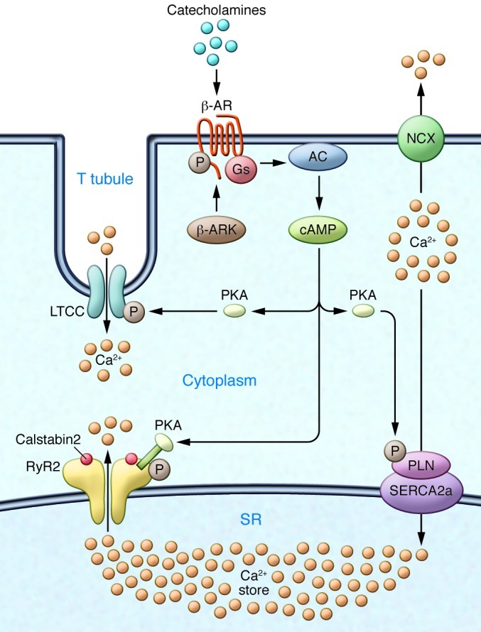
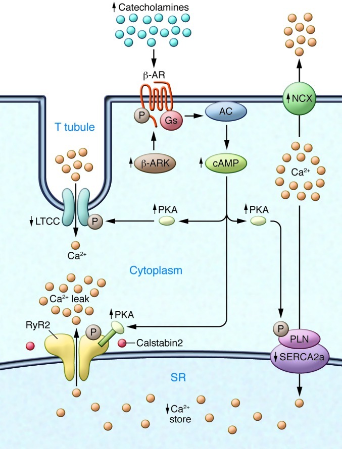
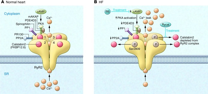
Ca2+-dependent signaling is highly regulated in cardiomyocytes and determines the force of cardiac muscle contraction. Ca2+ cycling refers to the release and reuptake of intracellular Ca2+ that drives muscle contraction and relaxation. In failing hearts, Ca2+ cycling is profoundly altered, resulting in impaired contractility and fatal cardiac arrhythmias. The key defects in Ca2+ cycling occur at the level of the sarcoplasmic reticulum (SR), a Ca2+ storage organelle in muscle. Defects in the regulation of Ca2+ cycling proteins including the ryanodine receptor 2, cardiac (RyR2)/Ca2+ release channel macromolecular complexes and the sarcoplasmic/endoplasmic reticulum Ca2+ ATPase 2a (SERCA2a)/phospholamban complex contribute to heart failure. RyR2s are oxidized, nitrosylated, and PKA hyperphosphorylated, resulting in "leaky" channels in failing hearts. These leaky RyR2s contribute to depletion of Ca2+ from the SR, and the leaking Ca2+ depolarizes cardiomyocytes and triggers fatal arrhythmias. SERCA2a is downregulated and phospholamban is hypophosphorylated in failing hearts, resulting in impaired SR Ca2+ reuptake that conspires with leaky RyR2 to deplete SR Ca2+. Two new therapeutic strategies for heart failure (HF) are now being tested in clinical trials: (a) fixing the leak in RyR2 channels with a novel class of Ca2+-release channel stabilizers called Rycals and (b) increasing expression of SERCA2a to improve SR Ca2+ reuptake with viral-mediated gene therapy. There are many potential opportunities for additional mechanism-based therapeutics involving the machinery that regulates Ca2+ cycling in the heart.


Figures

Figure 1
Figure 1. Ca2+ cycling in cardiomyocytes and regulation by PKA.
EC coupling in the heart starts with depolarization of the T tubule, which activates voltage-gated L-type Ca2+ channels (LTCCs) in the plasma membrane. Ca2+ influx via LTCCs triggers Ca2+ release from the SR via RyR2 (SR Ca2+ release channel). During systole, the free intracellular Ca2+ concentration increases ten-fold from ∼100 nM to ∼1 μM, which enables muscle contraction. The β-AR signaling pathway can increase the Ca2+ transient by activating the trigger (LTCC), release (RyR2), and uptake (SERCA/phospholamban [SERCA/PLN]) pathways. Catecholamine activation of β-ARs allows for the activation of adenylate cyclase (AC), mediated by specific G proteins (Gs), and the generation of cAMP, which in turn activates PKA. Relaxation occurs after intracellular Ca2+ is pumped out of the cytoplasm by SERCA2a, which is regulated by phospholamban. In addition, Ca2+ is extruded from the cell by the sarcolemmal NCX. RyR2 is a macromolecular complex comprised of four RyR2 monomers, PP1-spinophilin, PP2A-PR130, PKA-PDE4D3-mAKAP, calstabin2, CaMKII, and calmodulin. Calsequestrin regulates luminal SR free Ca2+, and junctin and triadin help maintain the integrity of the T tubule–SR junction. β-ARK, β-AR kinase.
Figure 2
Figure 2. Defective Ca2+ handling in failing hearts due to sympathetic overactivity.
Chronic activity of the sympathetic nervous system leads to phosphorylation of the β-AR, activation of β-AR kinase, and desensitization of β-ARs. The LTCC is also phosphorylated, and NCX expression is upregulated. An important contributor to impaired Ca2+ handling in HF is PKA hyperphosphorylation of RyR2. This leads to a higher sensitivity to Ca2+-induced Ca2+ release at low cytoplasmic Ca2+ concentrations, resulting in increased RyR2 open probability at low Ca2+ concentrations and a diastolic SR Ca2+ leak. The long-term effect of the diastolic Ca2+ leak is depletion of SR Ca2+ stores. SERCA2a expression and activity are decreased in HF, which is linked to phospholamban hypophosphorylation. In contrast, NCX expression and activity are upregulated in HF. Arrows indicate increased or decreased expression or activity in HF.
Figure 3
Figure 3. Dysfunctional RyR2 in failing hearts.
(A) The RyR2 macromolecular complex includes four identical RyR2 subunits (numerals 1–4 indicate the four monomers). Each RyR2 subunit binds one calstabin2 (also known as FKBP12.6) as well as mAKAP, to which PKA catalytic and regulatory subunits and PDE4D3 are bound; PP2A and its targeting protein PR130; and PP1 and its targeting protein sphinophilin (accessory molecules are only shown for one of the four RyR2 subunits, except calstabin2, which is shown for all four RyR2 subunits). The β-adrenergic signaling pathway can activate PKA through the second messenger cAMP. (B) In HF, PKA hyperphosphorylation of Ser2809, due to reduced PDE4D3, PP1, and PP2A levels in the RyR2 macromolecular complex, depletes calstabin2 from the RyR2 channel complex. The functional effect of these changes in the macromolecular composition of RyR2 is a pathological increase in Ca2+-dependent activation of RyR2 and depletion of SR Ca2+ stores, as well as functional uncoupling of RyR2 from their neighboring channels. Ca2+ leak due to abnormal RyR2 channel openings may be prevented by treatment with β-blockers (BBs), which interfere with the upstream β-AR signaling pathway, or with Rycals, which selectively increase the binding affinity of calstabin2 to PKA-phosphorylated and oxidized RyR2.

References

    1. Mathers CD, Loncar D. Projections of global mortality and burden of disease from 2002 to 2030. PLoS Med. 2006;3(11):e442. doi: 10.1371/journal.pmed.0030442. - DOI - PMC - PubMed
    1. Kamouh A, Francis GS. Kamouh A, Francis GS. Contemporary management and research directions in advanced heart failure: where are we going? Congest Heart Fail. 2011;17(5):241–247. doi: 10.1111/j.1751-7133.2011.00243.x. - DOI - PubMed
    1. Dube P, Weber KT. Congestive heart failure: pathophysiologic consequences of neurohormonal activation and the potential for recovery: part II. Am J Med Sci. 2011;342(6):503–506. doi: 10.1097/MAJ.0b013e3182327527. - DOI - PubMed
    1. Lompre AM, Hajjar RJ, Harding SE, Kranias EG, Lohse MJ, Marks AR. Ca2+ cycling and new therapeutic approaches for heart failure. Circulation. 2010;121(6):822–830. doi: 10.1161/CIRCULATIONAHA.109.890954. - DOI - PMC - PubMed
    1. Marx SO, et al. PKA phosphorylation dissociates FKBP12.6 from the calcium release channel (ryanodine receptor): defective regulation in failing hearts. Cell. 2000;101(4):365–376. doi: 10.1016/S0092-8674(00)80847-8. - DOI - PubMed

Publication types

MeSH terms