Review
. 2013 Jan;123(1):62-70.
doi: 10.1172/JCI63068. Epub 2013 Jan 2.

Regenerating new heart with stem cells

Affiliations
Review

Regenerating new heart with stem cells

Piero Anversa et al. J Clin Invest. 2013 Jan.

Retraction in

  • Regenerating new heart with stem cells.
    Anversa P, Kajstura J, Rota M, Leri A. Anversa P, et al. J Clin Invest. 2018 Dec 3;128(12):5676. doi: 10.1172/JCI126075. Epub 2018 Dec 3. J Clin Invest. 2018. PMID: 30507606 Free PMC article. No abstract available.

Abstract

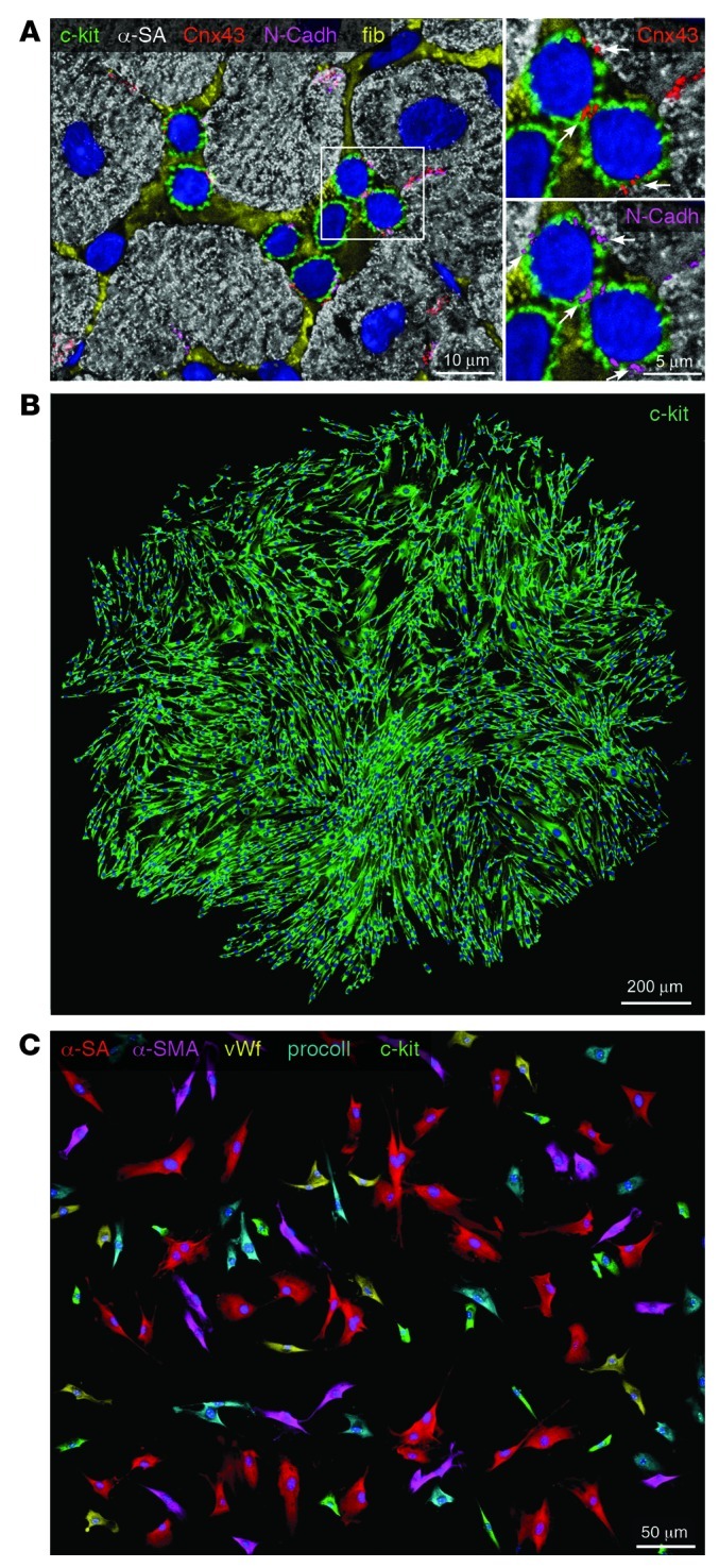
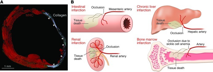
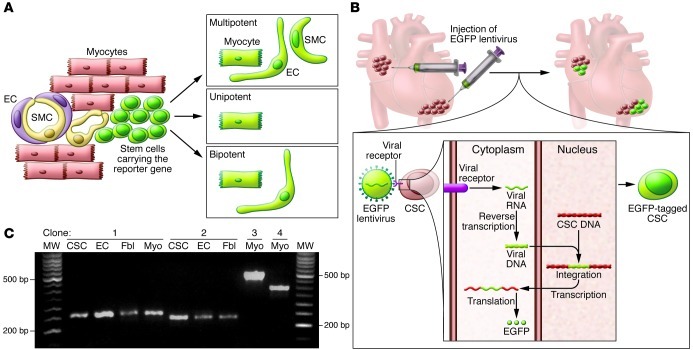
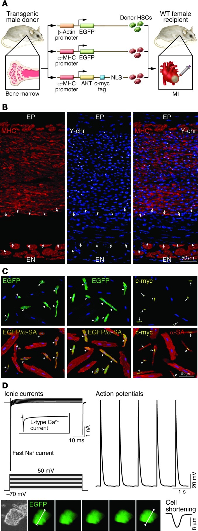
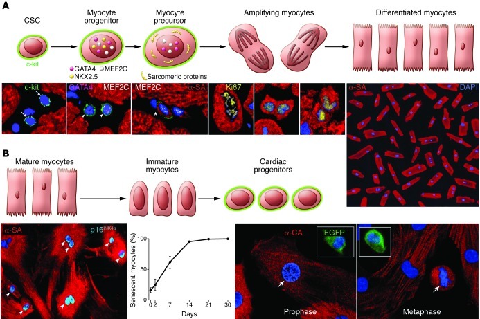
This article discusses current understanding of myocardial biology, emphasizing the regeneration potential of the adult human heart and the mechanisms involved. In the last decade, a novel conceptual view has emerged. The heart is no longer considered a postmitotic organ, but is viewed as a self-renewing organ characterized by a resident stem cell compartment responsible for tissue homeostasis and cardiac repair following injury. Additionally, HSCs possess the ability to transdifferentiate and acquire the cardiomyocyte, vascular endothelial, and smooth muscle cell lineages. Both cardiac and hematopoietic stem cells may be used therapeutically in an attempt to reverse the devastating consequences of chronic heart failure of ischemic and nonischemic origin.


Figures

Figure 1
Figure 1. Human CSCs.
(A) Cluster of c-kit–positive CSCs (green) surrounded by fibronectin (fib; yellow). Myocytes are labeled by α-sarcomeric actin (α-SA) (white). White rectangle indicates the area shown at higher magnification at right. Scale bar: 5 μm. Connexin 43 (Cnx43; red) and N-cadherin (N-Cadh; magenta) are expressed between c-kit–positive CSCs and between CSCs and cardiomyocytes (arrows). Scale bar: 10 μm. (B) Clone derived from deposition of a single c-kit–positive CSC in a well of a Terasaki plate. Cells in the clone are all c-kit positive (green). Scale bar: 200 μm. (C) In differentiating medium, clonal c-kit–positive CSCs differentiate into myocytes (α-SA, red), smooth muscle cells (α-smooth muscle actin [α-SMA]; magenta), endothelial cells (von Willebrand factor [vWf], yellow), and fibroblasts (procollagen [procoll], blue). Some undifferentiated c-kit–positive cells (green) are also present. Scale bar: 50 μm.
Figure 2
Figure 2. Infarcts in stem cell–regulated organs.
(A) Infarcted mouse heart characterized by collagen accumulation (white, arrowheads) in the region of healing. Myocytes are labeled by cardiac myosin heavy chain (MHC, red). Reproduced with permission from Proceedings of the National Academy of Sciences of the United States of America (ref. ; copyright 2001, National Academy of Sciences, USA). (B) Schematic representation of infarcted tissue in various organs. Regeneration of the infarct is absent in all cases.
Figure 3
Figure 3. Lineage tracing of CSCs.
(A) Representation of fate mapping involving the use of a fluorescent reporter gene (EGFP, green) driven by an inducible promoter coding for a stem cell–specific protein. Following differentiation and loss of stem cell antigen, myocytes, ECs, and SMCs continue to express EGFP, indicating a lineage relationship between ancestors and descendants. However, the labeled progeny may derive from activation of one or several stem cells, failing to document the multipotency of the parental stem cell. (B) Representation of viral gene tagging. Infection of CSCs with EGFP lentivirus results in the semirandom insertion of the proviral integrant in the genome of the recipient cell. Transcription and translation of the viral DNA result in expression of EGFP and fluorescent labeling of the infected CSCs. The unique insertion site of the viral genome is inherited by the entire population derived from the parental cell and can be amplified by PCR. CSCs nested in atrial and apical niches were labeled in situ to identify their progeny in vivo. (C) Four distinct clones were identified in EGFP-tagged CSCs, ECs, fibroblasts (Fbl), and cardiomyocytes (Myo) isolated from the ventricle of one mouse heart. Multiple PCR products (bands in agarose gel) were identified. Bands of the same molecular weight correspond to identical sites of integration of the proviral sequence in the host genome of CSCs, myocytes, ECs and fibroblasts, documenting the multipotency of CSCs in vivo. Reproduced with permission from Proceedings of the National Academy of Sciences of the United States of America (ref. ; copyright 2009, National Academy of Sciences, USA).
Figure 4
Figure 4. Transdifferentiation of c-kit–positive HSCs.
(A) Schematic representation of transgene constructs used in the generation of donor mice for the acquisition of HSCs to be delivered after infarction. In each case, the promoter that controls the ubiquitous (β-actin) or myocyte-restricted (α-MHC) expression of the transgene (EGFP or c-myc–tagged nuclear-targeted Akt) is shown. c-kit–positive HSCs from donor males were injected intramyocardially in wild-type female infarcted mice. NLS, nuclear localization signal (ref. 75). (B) Infarcted female mouse treated with male HSCs. Regenerated myocytes (left; MHC, red) carry the Y chromosome (center; Y-chr; white dots in nuclei). Merged image is shown at right. Arrows indicate non-regenerated infarct. A thin layer of spared myocytes is present in the epimyocardium (EP) and endomyocardium (EN) (ref. 75). Scale bar: 50 μm. (C) Examples of myocytes isolated from the regenerated myocardium of mice injected with HSCs collected from β-actin EGFP, α-MHC EGFP, or α-MHC c-myc–tagged nuclear-targeted Akt mice. Top panels illustrate the localization of EGFP (left and middle, green; arrowheads) and c-myc (right, yellow nuclei; arrows) in newly formed cardiomyocytes. Bottom panels show the colocalization of α-SA and EGFP (yellow, arrowheads) and of α-SA and c-myc (yellow nuclei, arrows). Large spared myocytes negative for EGFP or c-myc are also present (ref. 75). Scale bar: 50 μm. (D) Myocyte-specific ionic currents and action potentials of a cell isolated from the regenerated infarcted myocardium. EGFP-positive myocytes show contractile activity (bottom). Original magnification, ×200. Reproduced with permission from Proceedings of the National Academy of Sciences of the United States of America (ref. ; copyright 2007, National Academy of Sciences, USA).
Figure 5
Figure 5. Cellular mechanisms of myocardial homeostasis.
(A) Lineage relationship between CSCs and cardiomyocytes: CSC commitment results progressively in the formation of myocyte progenitors, myocyte precursors, and transit-amplifying myocytes, which divide and differentiate into mature myocytes. CSCs are c-kit–positive lineage-negative cells. Progenitors express c-kit and myocyte-specific transcription factors (GATA4, Nkx2.5, MEF2C) but do not show sarcomeric proteins. Precursors possess c-kit, myocyte-specific transcription factors, and sarcomeric proteins. Amplifying cells have myocyte nuclear and cytoplasmic proteins but are c-kit negative. This hierarchy is shown by immunolabeling and confocal microscopy (bottom). Micrographs from left to right show lineage-negative CSCs (arrows); myocyte progenitors (arrowheads) expressing c-kit, GATA4, and MEF2C; myocyte precursor (asterisk) showing c-kit, MEF2C, and α-SA; and three transit-amplifying myocytes (α-SA). Chromosomes are organized in anaphase-telophase and are Ki67 positive. Original magnification, ×700. Terminally differentiated cardiomyocytes from the human heart are shown at far right. Original magnification, ×100. Reproduced with permission from Proceedings of the National Academy of Sciences of the United States of America (ref. ; copyright 2003, National Academy of Sciences, USA). (B) The postulated process of dedifferentiation of postmitotic myocytes to immature myocytes and cardiac progenitors is represented schematically. Micrographs document that culture of differentiated myocytes is not coupled with reentry into the cell cycle and reversal to a primitive state. From left to right, adult cardiomyocytes in vitro show disassembly of the contractile apparatus and express the senescence-associated protein p16INK4a (bright blue, arrowheads; original magnification, ×250). The percentage of senescent myocytes increases with time. Fetal myocytes, isolated from a reporter mouse in which EGFP is driven by the c-kit promoter, divide in vitro (arrows) but do not express c-kit. Original magnification, ×500. Insets show c-kit–positive EGFP-positive CSCs used as a positive control. Reproduced with permission from Circulation Research (ref. 61).

References

    1. Rubart M, Field LJ. Cardiac regeneration: repopulating the heart. Annu Rev Physiol. 2006;68:29–49. doi: 10.1146/annurev.physiol.68.040104.124530. - DOI - PubMed
    1. Bergmann O, et al. Evidence for cardiomyocyte renewal in humans. Science. 2009;324(5923):98–102. doi: 10.1126/science.1164680. - DOI - PMC - PubMed
    1. Laflamme MA, Murry CE. Heart regeneration. Nature. 2011;473(7347):326–335. doi: 10.1038/nature10147. - DOI - PMC - PubMed
    1. Vunjak-Novakovic G, Lui KO, Tandon N, Chien KR. Bioengineering heart muscle: a paradigm for regenerative medicine. Annu Rev Biomed Eng. 2011;13:245–267. doi: 10.1146/annurev-bioeng-071910-124701. - DOI - PMC - PubMed
    1. Xu H, Yi BA, Chien KR. Shortcuts to making cardiomyocytes. Nat Cell Biol. 2011;13(3):191–193. doi: 10.1038/ncb0311-191. - DOI - PubMed