. 2013 Apr;33(4):524-31.
doi: 10.1038/jcbfm.2012.193. Epub 2013 Jan 2.

Hypoxia-inducible factor 1 is essential for spontaneous recovery from traumatic brain injury and is a key mediator of heat acclimation induced neuroprotection

Affiliations

Hypoxia-inducible factor 1 is essential for spontaneous recovery from traumatic brain injury and is a key mediator of heat acclimation induced neuroprotection

Gali Umschweif et al. J Cereb Blood Flow Metab. 2013 Apr.

Abstract

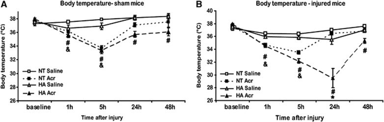
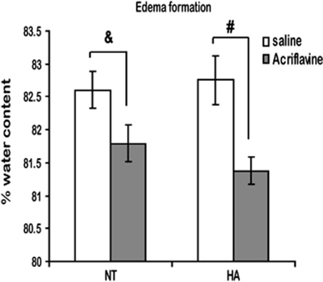
Heat acclimation (HA), a well-established preconditioning model, confers neuroprotection in rodent models of traumatic brain injury (TBI). It increases neuroprotective factors, among them is hypoxia-inducible factor 1α (HIF-1α), which is important in the response to postinjury ischemia. However, little is known about the role of HIF-1α in TBI and its contribution to the establishment of the HA protecting phenotype. Therefore, we aimed to explore HIF-1α role in TBI defense mechanisms as well as in HA-induced neuroprotection. Acriflavine was used to inhibit HIF-1 in injured normothermic (NT) or HA mice. After TBI, we evaluated motor function recovery, lesion volume, edema formation, and body temperature as well as HIF-1 downstream transcription targets, such as glucose transporter 1 (GLUT1), vascular endothelial growth factor, and aquaporin 4. We found that HIF-1 inhibition resulted in deterioration of motor function, increased lesion volume, hypothermia, and reduced edema formation. All these parameters were significantly different in the HA mice. Western blot analysis and enzyme-linked immunosorbent assay showed reduced levels of all HIF-1 downstream targets in HA mice, however, only GLUT1 was downregulated in NT mice. We conclude that HIF-1 is a key mediator in both spontaneous recovery and HA-induced neuroprotection after TBI.


Figures

Figure 1
Figure 1
Effects of acriflavine on hypoxia-inducible factor 1 (HIF-1) in the brain. (A) HIF-1β was immunoprecipitated (IP) with protein A beads and separated on SDS–polyacrylamide (PAGE) gels. Immunoblotting (IB) of HIF-1α indicates HIF-1 subunit dimerization in saline or acriflavine (Acr)-treated sham brain. (−) Immunoprecipitation of nonHIF-1α related protein (Bcl-xL) with the same beads. (+) Input from non immunoprecipitation blot of 60 μg protein from the same brain. (B) Injured tissue collected at 6 and 48 hours after injury or sham controls separated on SDS–PAGE gels and analyzed using western blotting. Optical density normalized to β-actin is shown. Quantified results are presented (#P<0.05 versus saline-treated mice of the matched time group, *P<0.05 versus NT mice in the sham of the saline-treated group, $P<0.05 versus sham mice in the saline-treated mice), determined by two-way ANOVA followed by Tukey post hoc tests, n=5 to 6 mice/group.
Figure 2
Figure 2
Acriflavine inhibits normothermic (NT) motor recovery after traumatic brain injury (TBI) and causes a deterioration in motor recovery of heat acclimation (HA) mice. Motor function was assessed and expressed using Neurological Severity Score (ΔNSS). The ΔNSS values were significantly higher in HA mice versus NT at 48 hours after injury (*P<0.05). Acriflavine (Acr) inhibited motor recovery of NT mice (⁁P<0.05 versus saline-treated mice), and induced deterioration of motor ability in HA mice at 24 hours and at 48 hours after injury (#P<0.05 versus saline-treated mice) determined by two-way ANOVA followed by Tukey post hoc tests, n⩾9 mice/group.
Figure 3
Figure 3
Acriflavine (Acr) increases lesion volume of injured heat acclimation (HA) mice. Lesion volume at 24 hours after injury was measured using 2,3,5-triphenyltetrazolium chloride (TTC) staining as the sum of the percentages of nonstained areas in different brain slices. Arrows point at the nonstained lesion area. The TTC staining quantification revealed significantly reduced lesion volumes in HA mice (*P<0.05). However, Acr significantly increased lesion volume in HA mice (#P<0.001) compared with NT mice, determined by one-way ANOVA n=8 to 12 mice/group.
Figure 4
Figure 4
Acriflavine (Acr) induces hypothermia. Baseline core body temperature was measured before closed head injury (CHI), as well as at 1, 5, 24, and 48 hours after Acr administration and sham operation (A) or CHI (B). Moderate hypothermia was measured shortly after sham operation both in normothermic (NT) and heat acclimation (HA) groups, however, only HA showed sustained hypothermia up to 48 hours after operation. Injury did not change temperature alternations induced by acriflavine alone (seen in NT mice), however, the temperature of acriflavine HA-treated mice declined significantly (#P<0.05 versus saline-treated HA mice, &P<0.05 versus saline-treated NT mice, and *P<0.001 versus sham HA mice of the matched group), determined by two-way ANOVA followed by Tukey post hoc tests. n=5/group for sham mice and n=9 to 32 mice/group for injured mice.
Figure 5
Figure 5
Acriflavine (Acr) reduces postinjury brain edema. Edema formation was determined 24 hours after closed head injury (CHI). Acriflavine reduced edema formation both in normothermic (NT) and in heat acclimation (HA) mice. (&P<0.01, #P<0.001) determined by one-way ANOVA. n=10 mice/group.
Figure 6
Figure 6
Acriflavine (Acr) alters protein levels of hypoxia-inducible factor 1 (HIF-1) downstream transcription targets. Extracts from injured tissue or sham controls were separated on SDS–PAGE gels and analyzed using western blotting. Optical density normalized to β-actin is shown. Quantified results are presented for 55kD GLUT1 (A), 45kD GLUT1 (B), and AQP4 (D). VEGF levels (C) were quantified using enzyme-linked immunosorbent assay (ELISA) (#P<0.05, respectively, to saline-treated mice of the same group, *P<0.05 versus normothermic (NT) mice of the matched treatment group, $P<0.05 versus sham mice of the matched group), determined by two-way ANOVA followed by Tukey post hoc tests. n=5 to 6 mice/group.

References

    1. Beit-Yannai E, Kohen R, Horowitz M, Trembovler V, Shohami E. Changes of biological reducing activity in rat brain following closed head injury: a cyclic voltammetry study in normal and heat-acclimated rats. J Cereb Blood Flow Metab. 1997;17:273–279. - PubMed
    1. Umschwief G, Shein NA, Alexandrovich AG, Trembovler V, Horowitz M, Shohami E. Heat acclimation provides sustained improvement in functional recovery and attenuates apoptosis after traumatic brain injury. J Cereb Blood Flow Metab. 2010;30:616–627. - PMC - PubMed
    1. Shein NA, Horowitz M, Alexandrovich AG, Tsenter J, Shohami E. Heat acclimation increases hypoxia-inducible factor 1alpha and erythropoietin receptor expression: implication for neuroprotection after closed head injury in mice. J Cereb Blood Flow Metab. 2005;25:1456–1465. - PubMed
    1. Leker RR, Shohami E. Cerebral ischemia and trauma-different etiologies yet similar mechanisms: neuroprotective opportunities. Brain Res Brain Res Rev. 2002;39:55–73. - PubMed
    1. Singh N, Sharma G, Mishra V. Hypoxia inducible factor-1: its potential role in cerebral ischemia. Cell Mol Neurobiol. 2012;32:491–507. - PMC - PubMed

MeSH terms

LinkOut - more resources