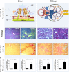
HDAC inhibitors in experimental liver and kidney fibrosis
- PMID: 23281659
- PMCID: PMC3564760
- DOI: 10.1186/1755-1536-6-1
HDAC inhibitors in experimental liver and kidney fibrosis
Abstract
Histone deacetylase (HDAC) inhibitors have been extensively studied in experimental models of cancer, where their inhibition of deacetylation has been proven to regulate cell survival, proliferation, differentiation and apoptosis. This in turn has led to the use of a variety of HDAC inhibitors in clinical trials. In recent years the applicability of HDAC inhibitors in other areas of disease has been explored, including the treatment of fibrotic disorders. Impaired wound healing involves the continuous deposition and cross-linking of extracellular matrix governed by myofibroblasts leading to diseases such as liver and kidney fibrosis; both diseases have high unmet medical needs which are a burden on health budgets worldwide. We provide an overview of the potential use of HDAC inhibitors against liver and kidney fibrosis using the current understanding of these inhibitors in experimental animal models and in vitro models of fibrosis.
Figures


Similar articles
-
HDAC Inhibitors: Therapeutic Potential in Fibrosis-Associated Human Diseases.Int J Mol Sci. 2019 Mar 16;20(6):1329. doi: 10.3390/ijms20061329. Int J Mol Sci. 2019. PMID: 30884785 Free PMC article. Review.
-
Comparison of the antifibrotic effects of the pan-histone deacetylase-inhibitor panobinostat versus the IPF-drug pirfenidone in fibroblasts from patients with idiopathic pulmonary fibrosis.PLoS One. 2018 Nov 27;13(11):e0207915. doi: 10.1371/journal.pone.0207915. eCollection 2018. PLoS One. 2018. PMID: 30481203 Free PMC article.
-
Anti-fibrotic effects of valproic acid: role of HDAC inhibition and associated mechanisms.Epigenomics. 2016 Aug;8(8):1087-101. doi: 10.2217/epi-2016-0034. Epub 2016 Jul 14. Epigenomics. 2016. PMID: 27411759 Review.
-
Epigenetic modifications by histone deacetylases: Biological implications and therapeutic potential in liver fibrosis.Biochimie. 2015 Sep;116:61-9. doi: 10.1016/j.biochi.2015.06.016. Epub 2015 Jun 25. Biochimie. 2015. PMID: 26116886 Review.
-
Blocking the class I histone deacetylase ameliorates renal fibrosis and inhibits renal fibroblast activation via modulating TGF-beta and EGFR signaling.PLoS One. 2013;8(1):e54001. doi: 10.1371/journal.pone.0054001. Epub 2013 Jan 16. PLoS One. 2013. PMID: 23342059 Free PMC article.
Cited by
-
The Emerging Role of MicroRNAs in NAFLD: Highlight of MicroRNA-29a in Modulating Oxidative Stress, Inflammation, and Beyond.Cells. 2020 Apr 22;9(4):1041. doi: 10.3390/cells9041041. Cells. 2020. PMID: 32331364 Free PMC article. Review.
-
Activation of Mir-29a in Activated Hepatic Stellate Cells Modulates Its Profibrogenic Phenotype through Inhibition of Histone Deacetylases 4.PLoS One. 2015 Aug 25;10(8):e0136453. doi: 10.1371/journal.pone.0136453. eCollection 2015. PLoS One. 2015. PMID: 26305546 Free PMC article.
-
The efficacy of vitamin E in preventing arthrofibrosis after joint replacement.Animal Model Exp Med. 2024 Apr;7(2):145-155. doi: 10.1002/ame2.12388. Epub 2024 Mar 25. Animal Model Exp Med. 2024. PMID: 38525803 Free PMC article.
-
Anti-fibrotic effects of valproic acid in experimental peritoneal fibrosis.PLoS One. 2017 Sep 5;12(9):e0184302. doi: 10.1371/journal.pone.0184302. eCollection 2017. PLoS One. 2017. PMID: 28873458 Free PMC article.
-
Epigenetic modifications of Klotho expression in kidney diseases.J Mol Med (Berl). 2021 May;99(5):581-592. doi: 10.1007/s00109-021-02044-8. Epub 2021 Feb 6. J Mol Med (Berl). 2021. PMID: 33547909 Review.
References
-
- Adam R, Karam V, Delvart V, O'Grady J, Mirza D, Klempnauer J, Castaing D, Neuhaus P, Jamieson N, Salizzoni M, Pollard S, Lerut J, Paul A, Garcia-Valdecasas JC, Rodríguez FS, Burroughs A. all contributing centers (www.eltr.org); European Liver and Intestine Transplant Association (ELITA) Evolution of indications and results of liver transplantation in Europe. A report from the European Liver Transplant Registry (ELTR) J Hepatol. 2012;57:675–688. doi: 10.1016/j.jhep.2012.04.015. - DOI - PubMed
-
- European Association for the Study of the Liver. EASL Annual Report 2009. European Association for the Study of the Liver, Geneva, Switzerland; 2010.
-
- Adam R, McMaster P, O'Grady JG, Castaing D, Klempnauer JL, Jamieson N, Neuhaus P, Lerut J, Salizzoni M, Pollard S, Muhlbacher F, Rogiers X, Garcia Valdecasas JC, Berenguer J, Jaeck D, Moreno Gonzalez E. European Liver Transplant Association. Evolution of liver transplantation in Europe: report of the European Liver Transplant Registry. Liver Transpl. 2003;9:1231–1243. doi: 10.1016/j.lts.2003.09.018. - DOI - PubMed
LinkOut - more resources
Full Text Sources
Other Literature Sources

