Review
. 2013 Mar;38(3):317-30.
doi: 10.3109/02713683.2012.747617. Epub 2013 Jan 2.

New twists to an old story: novel concepts in the pathogenesis of allergic eye disease

Affiliations
Review

New twists to an old story: novel concepts in the pathogenesis of allergic eye disease

Daniel R Saban et al. Curr Eye Res. 2013 Mar.

Abstract

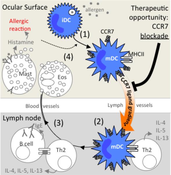
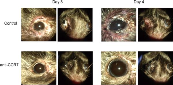
The prevalence of allergy is rising globally at a very significant rate, which is currently at 20-40% of individuals in westernized nations. In the eye, allergic conditions can take on the acute form such as in seasonal and perennial allergic conjunctivitis, or a more severe and debilitating chronic form such as in vernal and atopic keratoconjunctivitis. Indeed, some key aspects of allergic eye disease pathophysiology are understood, such as the role of mast cells in the acute allergic reaction, and the contribution of eosinophils in late-onset and chronic allergy. However, recent developments in animal models and clinical studies have uncovered new and important roles for previously underappreciated players, including chemokine receptors on ocular surface dendritic cells such as CCR7, the contribution of conjunctival epithelium to immunity, histamine and leukotriene receptors on conjunctival goblet cells and a role for mast cells in late-onset manifestations. Furthermore, recent work in animal models has delineated the contribution of IL-4 in the increased incidence of corneal graft rejection in hosts with allergic conjunctivitis. Recent studies such as these mean that conventional paradigms and concepts should be revisited. The aim of this review is to highlight some of the most recent advances and insights on newly appreciated players in the pathogenesis of allergic eye disease.


Figures

Figure 1
Figure 1
Conjunctival epithelial cell line (IOBA) expression of IL-17RD following exposure to TLR-2, -3, or -4 ligands (zymosan, Poly I:C and LPS, respectively), at 2, 6 and 24hr post treatment.
Figure 2
Figure 2
Presence of histamine receptors in rat conjunctiva and cultured goblet cells and stimulation of high molecular weight glycoconjugate secretion from cultured goblet cells. A. Rat conjunctiva and cultured goblet cells were homogenized and western blot analysis performed under denaturing conditions with anti-histamine receptor subtype antibodies. The blot is representative of three individual animals. H1R, histamine receptor subtype 1; H2R, histamine receptor subtype 2; H3R, histamine receptor subtype 3; H4R, histamine receptor subtype 4. B. Rat conjunctiva was removed and fixed. Rat conjunctival goblet cells were grown on glass slides and fixed. Immunofluorescence experiments were performed with antibodies against H3R. Magnification is 200 x. Micrographs are representative of three independent experiments. Arrows indicate the location of goblet cells. EP-epithelium. C. Effect of agonists specific to histamine receptor subtypes on goblet cell glycoconjugate secretion. Cultured goblet cells from rats were incubated for 2 h with the concentration of agonists that caused maximum secretion. The agonists used were histamine (10−5 M), the H1 receptor agonist histamine dimaleate (10−6 M), the H2 receptor agonist amthamine (10−4 M), the H3 receptor agonist α-methylhistamine (10−5 M), and the H4 receptor agonist 4-methylhistamine (10−5 M). Glycoconjugate secretion was measured by enzyme-linked lectin assay. Data are mean±SEM of seven independent experiments. * indicates statistical significance from no addition (B, basal). From Hayashi D et al. Invest Ophthalmol Vis Sci. 2012;53: 2993-3003; DOI:10.1167/iovs. 11-8748.
Figure 3
Figure 3
Identification of cysteinyl leukotriene receptors in rat conjunctival epithelium and cultured rat conjunctival goblet cells and stimulation of high molecular weight glycoconjugate secretion from cultured rat goblet cells. A. The presence of CysLT1 and CysLT2 receptors was determined by western blot analysis and is shown in upper panels. Each lane represents a separate animal. Fluorescence micrographs show localization of CysLT1 and CysLT2 receptors in conjunctiva (middle panels) and cultured goblet cells (lower panels). CysLT receptors are shown in red, and UEA-I, which indicates goblet cell secretory granules, is shown in green. Arrows indicate basolateral membranes of goblet cells. Arrowhead indicates stratified squamous cells. Original magnification X200. Micrographs are representative of three separate animals. B. Effect of leukotrienes on glycoconjugate secretion from cultured rat conjunctival goblet cells. Cultured rat conjunctival goblet cells were serum starved for 2 h before stimulation with LTC4 for 2 h (top left panel), LTD4 for 0.5 h (top right panel), or LTE4 for 1 h (bottom panel). The amount of glycoconjugate secretion was determined with ELLA. Data are mean ± SEM from four to five individual experiments. * p<0.05 from no addition, # p<0.01 from no addition (0). Open circle, the effect of carbachol used for 2 h. From: Dartt D et al J Immunol 2011; 186:4455-4466. doi: 10.4049/jimmunol.1000833
Figure 4
Figure 4
Hypothetical roles of vimentin in FcεRI- and CCR1-mediated signaling pathways in activated mast cells. FcεRI and CCR1 activation synergistically induces phosphorylation of ERK1/2, p38 MAP kinases and vimentin in activated mast cells. Disassembled vimentin filaments, unbundled by phosphorylation, may play a role in morphology and motility changes of activated mast cells. As disassembled vimentin fibers further interact with phosphorylated ERK1/2 and p38 MAPK, vimentin may also serve as a shuttle protein for nuclear entry of MAP kinases in mast cells.
Figure 5
Figure 5
Model proposed explaining the therapeutic effect of topical CCR7 blockade in allergic eye disease. (1) Exposed allergen to the ocular surface is captured by immature steady state DCs, which then begin to up-regulate CCR7 expression as they mature phenotypically (e.g. MHC II). This enables DCs to respond chemotactically to CCR7 ligands CCL19 and CCL21. (2) Increased expression of CCR7 ligands by lymphatic endothelial cells, LN subcapsular sinus, and high endothelial venules, directs DCs via lymphatic vessels to the LN paracortex for activation of Th2. (3) B cells further activate Th2 cells, which lead to secretion of Th2 cytokines (IL-4, IL-5, and IL-13) and consequent differentiation of B cells into IgE secreting plasma cells. (4) Mast cells can now ligate IgE, and their binding of allergen leads to the release of granules comprised of pro-inflammatory mediators such as histamine. Th2 cytokines cause eosinophils to release granules as well. This triggers the allergic reaction, as well maturation and infiltration of inflammatory DCs, which likewise enter into this feedback loop. Thus, the therapeutic effect of topical CCR7 blockade is thought to occur via inhibition DC homing and consequent Th2 activity.
Figure 6
Figure 6
Therapeutic effect of topical CCR7 antibody blockade in the mouse model of AC. Allergen primed mice were challenged topically with allergen once a day for 4 days to induce AC. Topical instillation of CCR7 blocking antibody (or isotype control) was given along with challenges. Representative images are shown from challenges on Day 3 and Day 4. Taken from Schlereth et al, Am J Pathol. 2012 Jun;180(6):2351-60.
Figure 7
Figure 7
Proposed mechanism whereby allergic conjunctivitis exacerbates corneal allograft rejection. A) CD4+CD25+ Tregs suppress CD4+ effector T cells by a contact-dependent mechanism involving TGF-β, CTLA-4, and glucocorticoid-induced tumor necrosis factor receptor (GITR) and thereby prevent corneal graft rejection. B) IL-4 produced during allergic conjunctivitis renders CD4+ effector T cells resistant to suppression by CD4+CD25+ Tregs.

References

    1. Leonardi A, Motterle L, Bortolotti M. Allergy and the eye. Clin Exp Immunol. 2008;153(Suppl 1):17–21. - PMC - PubMed
    1. Bielory L. Ocular allergy guidelines: a practical treatment algorithm. Drugs. 2002;62:1611–34. - PubMed
    1. Metz DP, Bacon AS, Holgate S, Lightman SL. Phenotypic characterization of T cells infiltrating the conjunctiva in chronic allergic eye disease. J Allergy Clin Immunol. 1996;98:686–96. - PubMed
    1. Maggi E, Biswas P, Del Prete G, et al. Accumulation of Th-2-like helper T cells in the conjunctiva of patients with vernal conjunctivitis. J Immunol. 1991;146:1169–74. - PubMed
    1. Calder VL, Jolly G, Hingorani M, et al. Cytokine production and mRNA expression by conjunctival T-cell lines in chronic allergic eye disease. Clin Exp Allergy. 1999;29:1214–22. - PubMed

MeSH terms

LinkOut - more resources