Pathogenic characteristics of three genotype II porcine reproductive and respiratory syndrome viruses isolated from China
- PMID: 23282224
- PMCID: PMC3616938
- DOI: 10.1186/1743-422X-10-7
Pathogenic characteristics of three genotype II porcine reproductive and respiratory syndrome viruses isolated from China
Abstract
Background: We examined differences in pathogenicity in pigs from China that had been experimentally infected with porcine reproductive and respiratory syndrome virus (PRRSV).
Methods: We compared pathogenic characteristics of a field isolate (GX-1/2008F), two PRRSV isolates (HN-1/2008, YN-1/2008) propagated in cells, and GX-1/2008F that had been propagated in cells (GX-1/2008). The clinical courses, along with humoral and cell-mediated responses, were monitored for 21 days post-infection (DPI). Animals were sacrificed and tissue samples used for gross pathological, histopathological and ultrastructure examination.
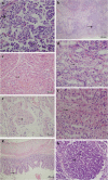
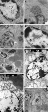
Results: At 2-3 DPI, animals infected with cell-propagated viruses exhibited signs of coughing, anorexia and fever. However their rectal temperature did not exceed 40.5°C. Viremia was detectable as early as 3 DPI in animals infected with HN-1/2008 and YN-1/2008. Animals inoculated with GX-1/2008F displayed clinical signs at 6 DPI; the rectal temperature of two animals in this group exceeded 41.0°C, with viremia first detected at 7 DPI. Seroconversion for all challenged pigs, except those infected with GX-1/2008, was seen as early as 7 DPI. All of these pigs had fully seroconverted by 11 DPI. All animals challenged with GX-1/2008 remained seronegative until the end of the experiment. Innate immunity was inhibited, with levels of IFN-α and IL-1 not significantly different between control and infected animals. The cytokines IFN-γ and IL-6 transiently increased during acute infection. All virus strains caused gross lesions including multifocal interstitial pneumonia and hyperplasia of lymph nodes. Inflammation of the stomach and small intestine was also observed. Lesions in the group infected with GX-1/2008F were more serious than in other groups. Transmission electron microscopy revealed that alveolar macrophages, plasmacytes and lymphocytes had fractured cytomembranes, and hepatocytes had disrupted organelles and swollen mitochondria.
Conclusions: The pathogenicity of the PRRSV field isolate became attenuated when propagated in MARC-145 cells. Tissue tropism of highly pathogenic strains prevailing in China was altered compared with classical PRRSV strains. The observed damage to immune cells and modulation of cytokine production could be mechanisms that PRRSV employs to evade host immune responses.
Figures


Similar articles
-
Genomic and pathogenicity analysis of two novel highly pathogenic recombinant NADC30-like PRRSV strains in China, in 2023.Microbiol Spectr. 2024 Oct 3;12(10):e0036824. doi: 10.1128/spectrum.00368-24. Epub 2024 Aug 20. Microbiol Spectr. 2024. PMID: 39162500 Free PMC article.
-
Immunopathological features of highly pathogenic Korean Lineage B PRRSV-2: insights into virulence indicators and host immune responses.Front Immunol. 2025 Jun 18;16:1599468. doi: 10.3389/fimmu.2025.1599468. eCollection 2025. Front Immunol. 2025. PMID: 40607395 Free PMC article.
-
Genomic sequence and virulence of a novel NADC30-like porcine reproductive and respiratory syndrome virus isolate from the Hebei province of China.Microb Pathog. 2018 Dec;125:349-360. doi: 10.1016/j.micpath.2018.08.048. Epub 2018 Aug 24. Microb Pathog. 2018. PMID: 30149129
-
The combination of PRRS virus and bacterial endotoxin as a model for multifactorial respiratory disease in pigs.Vet Immunol Immunopathol. 2004 Dec 8;102(3):165-78. doi: 10.1016/j.vetimm.2004.09.006. Vet Immunol Immunopathol. 2004. PMID: 15507303 Free PMC article. Review.
-
Host-pathogen interactions during porcine reproductive and respiratory syndrome virus 1 infection of piglets.Virus Res. 2015 Apr 16;202:135-43. doi: 10.1016/j.virusres.2014.12.026. Epub 2015 Jan 2. Virus Res. 2015. PMID: 25559070 Free PMC article. Review.
Cited by
-
Porcine cis-acting lnc-CAST positively regulates CXCL8 expression through histone H3K27ac.Vet Res. 2024 May 7;55(1):56. doi: 10.1186/s13567-024-01296-9. Vet Res. 2024. PMID: 38715098 Free PMC article.
-
Pathogenicity Studies of NADC34-like Porcine Reproductive and Respiratory Syndrome Virus LNSY-GY and NADC30-like Porcine Reproductive and Respiratory Syndrome Virus GXGG-8011 in Piglets.Viruses. 2023 Nov 11;15(11):2247. doi: 10.3390/v15112247. Viruses. 2023. PMID: 38005924 Free PMC article.
-
Porcine reproductive and respiratory syndrome virus (PRRSV) up-regulates IL-8 expression through TAK-1/JNK/AP-1 pathways.Virology. 2017 Jun;506:64-72. doi: 10.1016/j.virol.2017.03.009. Epub 2017 Mar 24. Virology. 2017. PMID: 28347884 Free PMC article.
-
Comparative pathogenesis of type 1 (European genotype) and type 2 (North American genotype) porcine reproductive and respiratory syndrome virus in infected boar.Virol J. 2013 May 21;10:156. doi: 10.1186/1743-422X-10-156. Virol J. 2013. PMID: 23687995 Free PMC article.
-
Susceptibility of porcine pulmonary microvascular endothelial cells to porcine reproductive and respiratory syndrome virus.J Vet Med Sci. 2020 Oct 7;82(9):1404-1409. doi: 10.1292/jvms.20-0324. Epub 2020 Aug 21. J Vet Med Sci. 2020. PMID: 32830156 Free PMC article.
References
-
- Keffaber KK. Reproductive failure of unknown etiology. Am Assoc Swine Veterinarians. 1989;10:1–10.
-
- Cavanagh D. Nidovirales: a new order comprising coronaviridae and arteriviridae. Arch Virol. 1997;10:629–633. - PubMed
Publication types
MeSH terms
Substances
LinkOut - more resources
Full Text Sources
Other Literature Sources

