. 2013 Apr 1;22(7):1358-72.
doi: 10.1093/hmg/dds546. Epub 2013 Jan 2.

Variable expressivity of ciliopathy neurological phenotypes that encompass Meckel-Gruber syndrome and Joubert syndrome is caused by complex de-regulated ciliogenesis, Shh and Wnt signalling defects

Affiliations

Variable expressivity of ciliopathy neurological phenotypes that encompass Meckel-Gruber syndrome and Joubert syndrome is caused by complex de-regulated ciliogenesis, Shh and Wnt signalling defects

Zakia A Abdelhamed et al. Hum Mol Genet. .

Abstract

The ciliopathies are a group of heterogeneous diseases with considerable variations in phenotype for allelic conditions such as Meckel-Gruber syndrome (MKS) and Joubert syndrome (JBTS) even at the inter-individual level within families. In humans, mutations in TMEM67 (also known as MKS3) cause both MKS and JBTS, with TMEM67 encoding the orphan receptor meckelin (TMEM67) that localizes to the ciliary transition zone. We now describe the Tmem67(tm1(Dgen/H)) knockout mouse model that recapitulates the brain phenotypic variability of these human ciliopathies, with categorization of Tmem67 mutant animals into two phenotypic groups. An MKS-like incipient congenic group (F6 to F10) manifested very variable neurological features (including exencephaly, and frontal/occipital encephalocele) that were associated with the loss of primary cilia, diminished Shh signalling and dorsalization of the caudal neural tube. The 'MKS-like' group also had high de-regulated canonical Wnt/β-catenin signalling associated with hyper-activated Dishevelled-1 (Dvl-1) localized to the basal body. Conversely, a second fully congenic group (F > 10) had less variable features pathognomonic for JBTS (including cerebellar hypoplasia), and retention of abnormal bulbous cilia associated with mild neural tube ventralization. The 'JBTS-like' group had de-regulated low levels of canonical Wnt signalling associated with the loss of Dvl-1 localization to the basal body. Our results suggest that modifier alleles partially determine the variation between MKS and JBTS, implicating the interaction between Dvl-1 and meckelin, or other components of the ciliary transition zone. The Tmem67(tm1(Dgen/H)) line is unique in modelling the variable expressivity of phenotypes in these two ciliopathies.


Figures
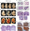
Figure 1.
Figure 1.
MKS-like neurodevelopmental defects in Tmem67−/− mouse embryos: (A) whole embryo images showing phenotypic variability in Tmem67−/− compared with Tmem67+/+ congenic littermates at embryonic age E11.5. Indicated are occipital meningocele (arrows), cranial neural tube defects affecting midbrain and hindbrain regions, localized areas of spinal neural tube defects (braces) and cystic dilated fourth ventricle and expanded roof plate (arrowhead). Scale bar = 5 mm. (B) H&E stained horizontal brain sections of E11.5 Tmem67−/− mutants. Top panels: meningocele with a prominent defect in the head mesoderm (arrowhead) surrounding the hindbrain, dilated fourth ventricle (4V) and roof plate malformation (asterisk). Bottom panel: failure of neural groove closure at the level of the mesencephalon (arrowhead). Scale bars = 1 mm. (C) Whole embryo images of E11.5 congenic littermates showing anterior neuropore closure defect (forebrain haemorrhage) (middle panel, arrowhead), and midbrain exencephaly (middle panel, arrow) and forebrain exencephaly (right panel, arrowhead) in Tmem67−/−. (D) H&E sections of occipital encephalocele (arrowheads and asterisk) in E15.5 Tmem67−/− embryos. Scale bar = 0.1 mm. (E) Frontal encephalocele in E15.5 Tmem67−/− embryos (arrowheads, detail shown in insets). (F) Semi-lobar holoprosencephaly in P0 Tmem67−/− pups showing fusion of the two lateral ventricles (asterisk), absence of the anterior commissar (ac) and corpus callosum (cc) dysgenesis (indicated by braces), enlarged hippocampus (hp) and basal ganglia (bg), reduced cortical thickness and frontal encephalocele (arrowhead). (G) E18.5 Tmem67−/− brain sectioned and stained for activated β-catenin and the neuronal differentiation marker Tuj1. Arrowheads indicate a region of neuronal progenitors in a frontal encephalocele (asterisk). Scale bar = 1 mm.
Figure 2.
Figure 2.
JBTS-like posterior fossa and cerebellar hypoplasia defects in Tmem67−/− mouse embryos: (A, left panels) small hind brain region in E11.5 Tmem67−/− embryos and smaller forebrain (asterisk). (Middle panel) Reduced hind brain region (brace) and hypoplastic mandible (arrowhead) in P0 Tmem67−/− pups. (Right panel) Reduced anteroposterior axis (dotted lines) of the developing forebrain in E15.5 Tmem67−/− embryos, quantitated in the bar graph. Values shown are means of three independent replicates. (B) Horizontal brain sections of E15.5 Tmem67−/− embryos showing pathognomonic features of JBTS, including a deep interpeduncular fossa (top panel, arrowhead) and reduction in the anteroposterior axis of the midbrain, and cerebellar vermis aplasia or hypoplasia (middle and bottom panels, asterisk). (C) Mid-sagittal sections of Tmem67−/− E12.5 embryos showing cerebellar vermis hypoplasia. Scale bars = 50 μm. (D) Complex posterior fossa defects in the E15.5 Tmem67−/− embryonic hindbrain (top panel, with middle panel showing magnified insets indicated by black frames), showing marked dysplasia of the caudal medulla, absence of the inferior olive nucleus (dashed oval, arrow in mutant), deep abnormal interpeduncular fossa (black asterisk), abnormally dilated cisterna magna and septa (arrowhead) and an enlarged and protruding locus coeruleus (red asterisk). (Lower panel) Dysplastic cervico-medullary junction (arrowhead) and overlying head structure (asterisk). Scale bar = 1 mm. Abbreviations: 4V, fourth ventricle; aq, aquaduct; cb, cerebellum; cbh, cerebellar hemispheres; cv, cerebellar vermis; ic, inferior colliculus; ion, inferior olive nucleus ipf, interpenduncular fossa.
Figure 3.
Figure 3.
Different cilia defects in Tmem67−/− embryos with MKS-like and JBTS-like phenotypes: (A, top panels) Horizontal section of the developing E11.5 caudal neural tube in litter-mate Tmem67+/+ and ‘MKS-like’ Tmem67−/− embryo at the level of the anterior limb buds stained for acetylated α-tubulin (red), centrosomes/basal bodies (γ-tubulin, green) and nuclei (DAPI, blue). (Bottom panels) Magnified insets (white frames) with primary cilia in the Tmem67+/+ neural tube indicated by arrowheads. Scale bar = 10 μm. (B) E14.5 neuroepithelium of the lateral wall of the third ventricle (3V) in litter-mate Tmem67+/+ and ‘MKS-like’ Tmem67−/− embryo stained as for (A). Scale bar = 10 μm. (C) SEM images of E18.5 lateral ventricles showing loss of primary cilia and disruption in the planar polarization of the ependymal cell layer in an ‘MKS-like’ Tmem67−/− embryo. Magnified insets (orange frames) show detail of cilia (arrowheads) in a littermate Tmem67+/+ embryo. Scale bar = 50 μm with 10 μm subdivisions. (D, top panels) Horizontal sections as in (A) for littermate Tmem67+/+ and ‘JBTS-like’ Tmem67−/− embryo stained for acetylated α-tubulin (red), centrosomes/basal bodies (γ-tubulin, green) and nuclei (DAPI, blue). (Bottom panels) Magnified insets (white frames) with primary cilia indicated by arrowheads. Scale bar = 10 μm. (E) IF microscopy (top panels) and SEM images (bottom panels) of E18.5 lateral ventricles showing the presence of abnormal, bulbous cilia and defective planar organization of the ependymal layer in ‘JBTS-like’ Tmem67−/− embryos. Magnified insets (orange frames) show detail of cilia (arrowheads). Scale bar = 50 μm with 10 μm subdivisions.
Figure 4.
Figure 4.
Defective dorsoventral patterning and Shh signalling in Tmem67−/− mutants: (A) loss of Shh signalling and dorsalization of the caudal neural tube in ‘MKS-like’ embryos. (Top panel) Shh protein expression (red) at the notochord (nc, arrowheads) and induced expression at the floor plate (FP) in the Tmem67+/+ embryo. (Bottom panels) IHC staining for the indicated dorsoventral patterning markers and haemotoxylin counterstaining. Note the variable thinning of the roof plate (RP) in Tmem67−/− embryos. Scale bar = 100 μm. (B) Activation of the Shh signalling pathway and mild ventralization defects in ‘JBTS-like’ embryos using IHC staining for the indicated dorsoventral patterning markers. Note the absence of any roof plate defect in Tmem67−/− embryos.
Figure 5.
Figure 5.
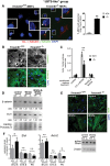
In vivo and in vitro up-regulated canonical Wnt/β-catenin signalling defects in Tmem67−/− embryos with MKS-like phenotypes: (A, top panels) IF confocal microscopy of horizontal midbrain sections of E11.5 embryos, with regions magnified in the bottom panels indicated by white frames in top panels. The Tmem67−/− mutant shows a prominent meningocele (bottom panel, asterisk), stained for the primary ciliary axoneme (acetylated α-tubulin, red), activated β-catenin (green) and nuclei (DAPI, blue). Magnified insets (white frames) show detail of primary cilia (arrowheads) in the Tmem67+/+ embryo; 4V, fourth ventricle. Scale bar = 20 μm. (B) IF microscopy of Tmem67+/+ MEFs and Tmem67−/− MEFs derived from embryos with an MKS-like phenotype, stained for acetylated α-tubulin (red), centrosomes/basal bodies (γ-tubulin, green) and nuclei (DAPI, blue). Magnified insets (white frames) show detail of ciliogenesis and centrosomal defects in Tmem67−/− MEFs. Three separate MEF lines for both genotypes were derived and consistently assayed at passage 3. Bar graphs quantify ciliogenesis (presence of a cilium defined as a single, uniform axoneme > 2 μm in length; absence defined as <1 μm or complete failure of ciliogenesis) and centrosomal/basal body defects (>5 μm separation, or multiple centrosomes/dispersal of pericentriolar material). Scale bars = 20 μm. (C) Immunoblotting of whole cell lysates extracted from wild-type Tmem67+/+ (+/+), heterozygous Tmem67+/− (+/−) and mutant ‘MKS-like’ Tmem67−/− (−/−) MEFs for Dishevelled-1 (Dvl1, phosphorylated P-Dvl1 also indicated), total β-catenin and cyclin D1. Immunoblotting for β-actin is the loading control. The ratio of β-catenin band intensity: loading control band intensity is indicated. (D) Whole cell lysates from MEFs of the indicated genotypes stimulated with Wnt3a-conditioned media immunoblotted for phosphorylated (P)-Lrp6 and total β-catenin. The ratio of β-catenin band intensity: loading control band intensity (‘ratio’) is indicated below the β-catenin panel. (E) Whole cell lysates from MEFs of the indicated genotypes treated with Wnt5a and assayed for active RhoA levels by a pulldown assay. (F) Bar graph on left: TOPFlash assays for Tmem67+/+ (grey) and ‘MKS-like’ Tmem67−/− (black) MEFs following treatment with the indicated conditioned media. Bar graph on right: dysregulated canonical Wnt signalling in ‘MKS-like’ Tmem67−/− cells following Wnt3a treatment is rescued by cotransfection with wild-type HA-TMEM67 expression construct but not empty vector control. Values shown are means of at least four independent replicates.
Figure 6.
Figure 6.
Down-regulated canonical Wnt/β-catenin signalling in Tmem67−/− embryos with JBTS-like phenotypes: (A, left panel) IF confocal microscopy of Tmem67+/+ MEFs and Tmem67−/− MEFs derived from embryos with a JBTS-like phenotype, stained for acetylated α-tubulin (red), γ-tubulin (green) and nuclei (blue). Magnified insets (white frames) show detail of cilia. (Right panel) The bar graph quantifies the number of abnormally long cilia (>5 μm). Scale bar = 20 μm. (B) MEFs stimulated with Wnt3a and stained for β-catenin (green) and nuclei (blue) show normal nuclear translocation of β-catenin in Tmem67+/+ MEFs (arrowheads) but not in Tmem67−/− MEFs. (C) TOPFlash assays in Tmem67+/+ and ‘JBTS’ Tmem67−/− MEFs following treatment with the indicated conditioned media. Values shown are means of at least four independent replicates. (D) Whole cell lysates extracted from wild-type Tmem67+/+ (+/+) and mutant ‘JBTS-like’ Tmem67−/− (−/−) MEFs, following treatment with the indicated conditioned media, were immunoblotted for β-catenin and Dishevelled-1 (Dvl-1), with Ponceau S staining as the loading control. The ratios of either β-catenin or Dvl-1 band intensities: loading control band intensity (‘ratio’) are indicated beneath the appropriate panel. (E, top panel) IF confocal microscopy of Tmem67+/+ and ‘JBTS’ Tmem67−/− MEFs for phalloidin to visualize F-actin following treatment with either control- or Wnt5a-conditioned media as indicated. (Bottom panel) Whole cell lysates from MEFs of the indicated genotypes treated with Wnt5a and assayed for active RhoA levels. (F) qRT-PCR results for Tmem67+/+ (grey) and JBTS-like Tmem67−/− (black) cortical tissue at embryonic ages E12.5, E15.5 and P0 as indicated, showing the expression levels of Shh (left panel) and Axin2 (right panel). Values shown are means of three independent biological replicates.
Figure 7.
Figure 7.
Disruption of the TMEM67/meckelin-Dishevelled-1 axis is associated with the JBTS-like phenotype. (A) IF confocal microscopy of Tmem67+/+ MEFs and Tmem67−/− MEFs derived from embryos with an MKS-like phenotype, stained for Dvl-1 (red), γ-tubulin (green) and and nuclei (blue). Arrowheads indicate the cells magnified in the insets below. Scale bar = 10 μm. (B) Tmem67+/+ MEFs and Tmem67−/− MEFs derived from embryos with a JBTS-like phenotype, stained as in (A). Scale bar = 10 μm. (C) Immunoprecipitation (IP) of endogenous Dvl-1 by wild-type TMEM67/meckelin. HEK293 cells were transfected with empty vector negative control (control), wild-type HA-meckelin (wt) or p.F919del mutant HA-meckelin (919delF) constructs. Expression of HA-tagged proteins has been determined previously by immunoblotting with anti-HA (Fig. 2C, ref. 26). (Left panel) 10% of total input WCE for each IP is indicated. (Right panel) Affinity-purified ‘Ct Ab’ anti-meckelin antibody against the C-terminus, and anti-HA rabbit polyclonal (Rb Abs) against epitope-tagged wild-type HA-meckelin, preferentially pull-down endogenous Dvl-1. Expression of the p.F919del mutant HA-meckelin (919delF) abrogated or prevented interaction with Dvl-1. IP for an irrelevant monoclonal antibody (irr. Ab.) did not pull-down Dvl-1.

References

    1. Alexiev B.A., Lin X., Sun C.-C., Brenner D.S. Meckel-Gruber syndrome: pathologic manifestations, minimal diagnostic criteria, and differential diagnosis. Arch. Pathol. Lab. Med. 2006;130:1236–1238. - PubMed
    1. Salonen R., Paavola P. Meckel syndrome. J. Med. Genet. 1998;35:497–501. - PMC - PubMed
    1. Ahdabbarmada M., Claassen D. A distinctive triad of malformations of the central-nervous-system in the Meckel-Gruber syyndrome. J. Neuropathol. Exp. Neurol. 1990;49:610–620. - PubMed
    1. Paetau A., Salonen R., Haltia M. Brain pathology in the Meckel syndrome—a study of 59 cases. Clin. Neuropathol. 1985;4:56–62. - PubMed
    1. Baala L., Romano S., Khaddour R., Saunier S., Smith U.M., Audollent S., Ozilou C., Faivre L., Laurent N., Foliguet B., et al. The Meckel-Gruber syndrome gene, MKS3, is mutated in Joubert syndrome. Am. J. Hum. Genet. 2007;80:186–194. - PMC - PubMed

Publication types

MeSH terms

Supplementary concepts