. 2013 Jan 2;33(1):93-105.
doi: 10.1523/JNEUROSCI.3841-12.2013.

Functional differentiation of a population of electrically coupled heterogeneous elements in a microcircuit

Affiliations

Functional differentiation of a population of electrically coupled heterogeneous elements in a microcircuit

Kosei Sasaki et al. J Neurosci. .

Erratum in

  • J Neurosci. 2013 Feb 6;33(6):2728. Sasaki, Kosai [corrected to Sasaki, Kosei]

Abstract

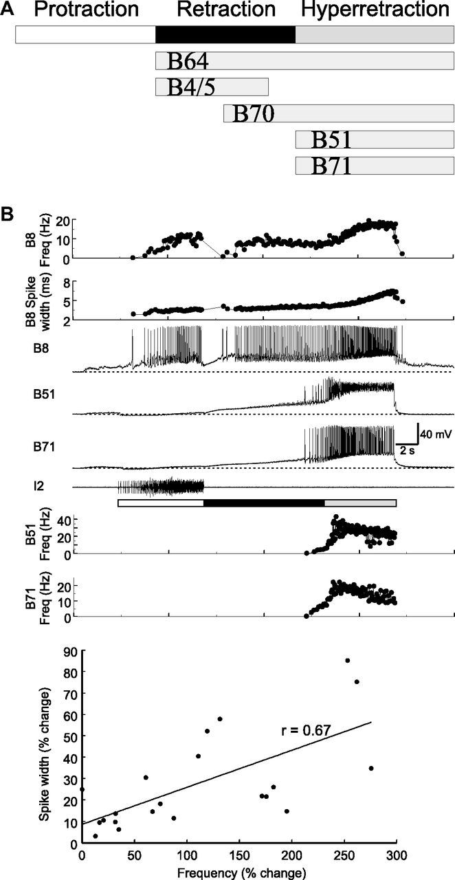
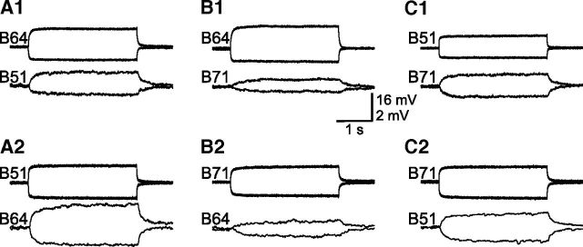
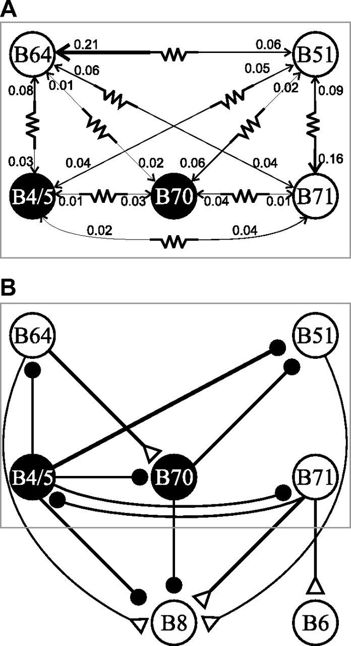
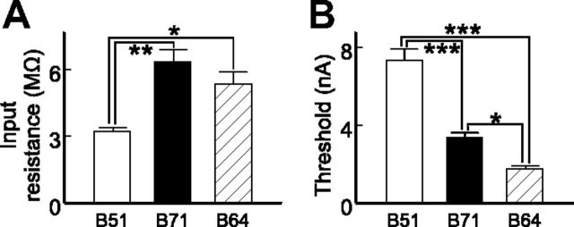
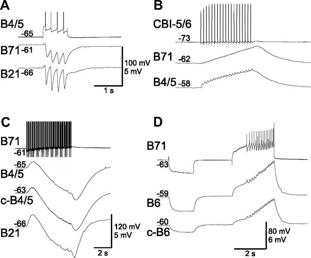
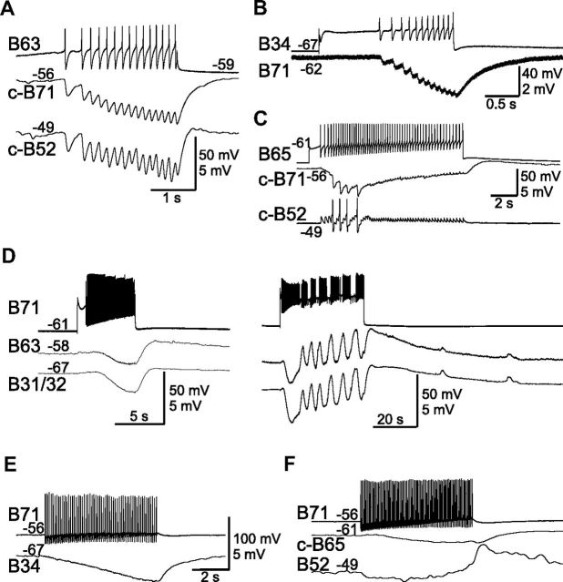
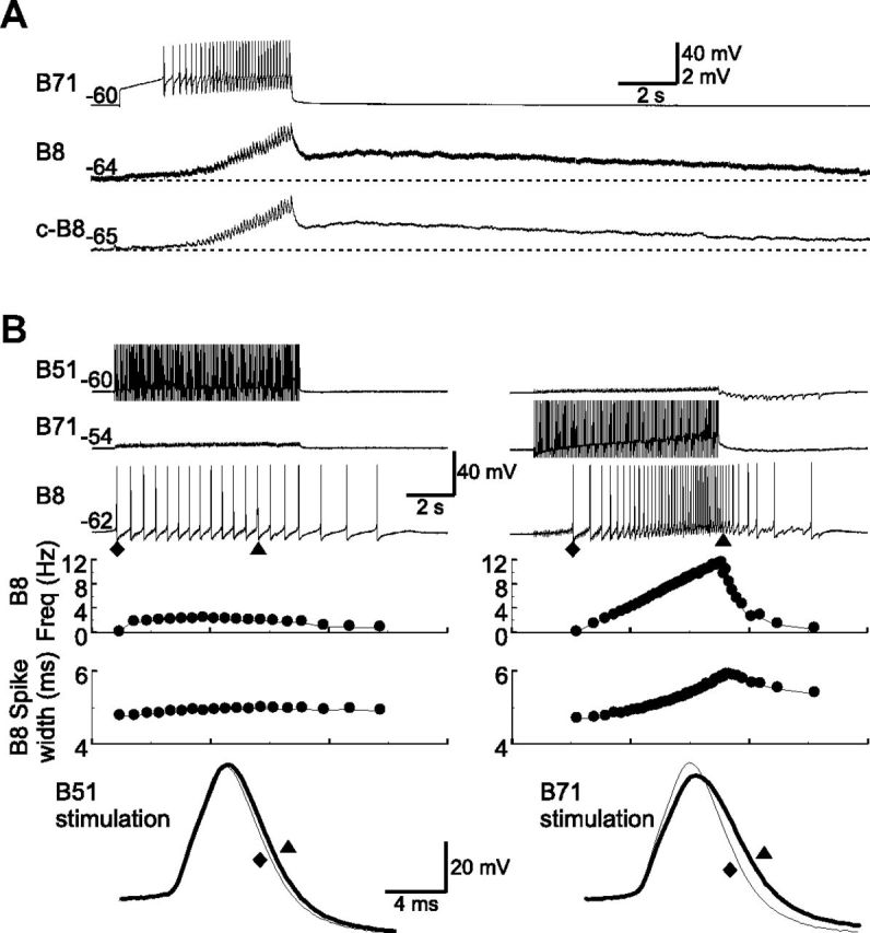
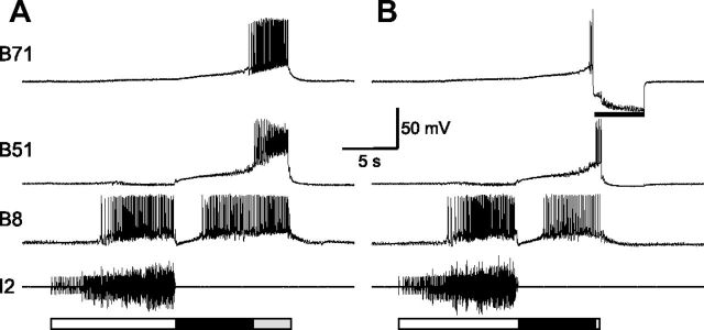
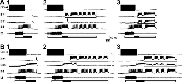
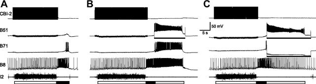
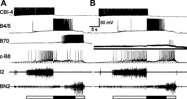
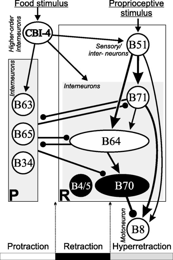
Although electrical coupling is present in many microcircuits, the extent to which it will determine neuronal firing patterns and network activity remains poorly understood. This is particularly true when the coupling is present in a population of heterogeneous, or intrinsically distinct, circuit elements. We examine this question in the Aplysia californica feeding motor network in five electrically coupled identified cells, B64, B4/5, B70, B51, and a newly identified interneuron B71. These neurons exhibit distinct activity patterns during the radula retraction phase of motor programs. In a subset of motor programs, retraction can be flexibly extended by adding a phase of network activity (hyper-retraction). This is manifested most prominently as an additional burst in the radula closure motoneuron B8. Two neurons that excite B8 (B51 and B71) and one that inhibits it (B70) are active during hyper-retraction. Consistent with their near synchronous firing, B51 and B71 showed one of the strongest coupling ratios in this group of neurons. Nonetheless, by manipulating their activity, we found that B51 preferentially acted as a driver of B64/B71 activity, whereas B71 played a larger role in driving B8 activity. In contrast, B70 was weakly coupled to other neurons and its inhibition of B8 counteracted the excitatory drive to B8. Finally, the distinct firing patterns of the electrically coupled neurons were fine-tuned by their intrinsic properties and the largely chemical cross-inhibition between some of them. Thus, the small microcircuit of the Aplysia feeding network is advantageous in understanding how a population of electrically coupled heterogeneous neurons may fulfill specific network functions.


Figures

Figure 1.
Figure 1.
Activity phasing of neuronal elements within the retraction circuit. A, Schematic representations of activity phasing of neurons in a single cycle. Each cycle of a motor program consists of protraction (open bar) followed by retraction (filled bar). In a subset of programs, as illustrated here, hyper-retraction (gray bar) is added on to the retraction phase. Five retraction neurons, B64, B4/5, B70, B51, and B71, show various activity patterns during retraction and hyper-retraction. B, In a single cycle of a program elicited by CBI-4 (data not shown), B51 and B71 showed similar activity patterns during hyper-retraction, accompanied by strong firing in B8. The bottom graph shows that increasing firing frequency (% change) and spike width (% change) of B8 during hyper-retraction (n = 21) are positively correlated. Line, Linear regression line; r, correlation coefficient. Protraction (open bar) is marked by I2 activity. Freq, Frequency.
Figure 2.
Figure 2.
Electrical coupling between three neuronal elements (B64, B51, and B71) within the retraction circuit. A1, A2, From B64 (both depolarizing and hyperpolarizing currents, 1.5 nA) to B51 (A1), and from B51 (3 nA) to B64 (A2); n = 4. B1, B2, From B64 (1.5 nA) to B71 (B1), and from B71 (2 nA) to B64 (B2); n = 3. C1, C2, From B51 (2 nA) to B71 (C1), and from B71 (2 nA) to B51 (C2); n = 4. For each panel, the top recordings show the presynaptic cells that were depolarized and hyperpolarized for 3 s, and the bottom recordings show corresponding responses in the postsynaptic cells. Coupling ratios measured by depolarizing and hyperpolarizing pulses were similar, suggesting that these electrical synapses are nonrectifying. Note the different coupling strengths between the coupled pairs and asymmetrical coupling within the coupled pairs. Membrane potentials at the beginning of traces were as follows: B64, −62 mV; B51, −57 mV; B71, −55 mV.
Figure 3.
Figure 3.
The two B71 neurons are both located in the right side of the buccal ganglion. A, Photograph of the buccal ganglion shows that B71, a bipolar interneuron, projects two axons bilaterally to the CBCs. B71 was injected with Alexa 488 dye. B, B71 soma area in higher magnification. C–H, The second B71 was injected with Alexa 568 dye. C, D, similar to A and B, except they were from the second B71. F, G, Photographs of the right and left cerebral ganglion around the roots of the CBCs, showing the second B71 projections that enter the cerebral ganglion. E, H, Higher-magnification views of the B71 axon terminals from the photographs in F and G, respectively. Buccal ganglion (rostral side up): BN, buccal nerve; EN, esophageal nerve; RN, radula nerve. Cerebral ganglion (ventral side up): AT, anterior tentacular nerve; ULAB, upper labial nerve; LLAB, lower labial nerve. CPC, Cerebral-pedal connective.
Figure 4.
Figure 4.
Schematic diagrams of synaptic connections of retraction circuitry neurons. A, Electrical coupling showing the coupling ratios. Thickness of lines is proportional to coupling ratios. B, Chemical connections. Also illustrated are connections to motoneurons B8 and B6. Two neurons, B4/5 and B70, that inhibit B8 are shown in filled circles. Open triangles, Chemical excitation; closed circles, inhibition; resistor symbol, electrical coupling.
Figure 5.
Figure 5.
Intrinsic properties of B51, B71, and B64. A, Apparent input resistances (n = 6). B, Spike thresholds are the minimum depolarizing currents needed to induce spiking (n = 4). *p < 0.05; **p < 0.01; ***p < 0.001 (Bonferroni post hoc tests). Error bars indicate SEM.
Figure 6.
Figure 6.
Chemical synaptic connections of B71 with other retraction neurons. A, B4/5 elicits one-for-one IPSPs in B71. Also shown is the B21 neuron, which is also inhibited. B, Retraction neuron CBI-5/6 excites B71 and B4/5. C, B71 inhibits B4/5 and B21. No unitary IPSPs can be resolved. D, B71 is electrically coupled to motoneuron B6. Spikes in B71 elicit large changes in B6 membrane potential, suggesting that B71 may also chemically excite B6. “c-” before cell names in this and the rest of the figures means “contralateral. ” Numbers at the beginning of traces are membrane potentials (in millivolts).
Figure 7.
Figure 7.
Synaptic connections of B71 with protraction neurons. A–C, Protraction interneurons B63 (A), B34 (B), and B65 (C) inhibit B71. A, B, B63 and B34 elicit one-for-one IPSPs in B71. B63 also inhibits B52. C, B65 also elicits one-for-one EPSPs in B52, and induces three spikes in B52 that in turn elicit three large-amplitude IPSPs in B71. D–F, B71 in turn inhibits B63 and B31/32 (D), B34 (E), B65 (F), and no unitary IPSPs can be resolved. In addition to the IPSPs, B71 also elicits a slow EPSP in B63 and B31/32 that is more obvious when B71 is stimulated for prolonged periods (D, right). Recording in B–E were made in high-divalent saline. Numbers at the beginning or the end of traces are membrane potentials (in millivolts).
Figure 8.
Figure 8.
Connections from B71 to radula closure motoneuron B8. A, B71 elicits fast and slow EPSPs in B8 (n = 25). The unitary EPSPs are facilitating and become more obvious as B71 stimulation progresses. Dotted line, Resting potentials of B8. Recordings were made in high-divalent saline. B, Comparisons of the excitatory actions from B51 (left, 20 Hz) and B71 (right, 15 Hz) to B8 (n = 4). The B71 effect is stronger. Note that when B71, but not B51, is stimulated, B8 firing frequency (Freq) increases progressively, and B8 spikes broaden. Bottom, Enlarged records of B8 show individual spikes of the first one (diamonds, thin lines) and the one with the longest spike width (triangles, thick lines) upon B51 (left) or B71 (right) stimulation. Numbers at the beginning of traces are membrane potentials (in millivolts).
Figure 9.
Figure 9.
Necessity of B71 for hyper-retraction. A, A single cycle of a motor program induced by CBI-4 stimulation. CBI-4 is not shown. B, Another cycle of a program elicited by CBI-4, and hyperpolarization of B71 (bar, −9 nA) prevents expression of hyper-retraction (n = 6).
Figure 10.
Figure 10.
B71 stimulation in motor programs elicited by CBI-4 does not recruit continued firing of B51 or B64. A1, B1, A single cycle of a motor program elicited by CBI-4 without hyper-retraction. A2, B2, Activation of B71 (bars) induces strong firing in B8 reminiscent of B8 firing in hyper-retraction, and neither B51 (A2; n = 3) nor B64 (B2; n = 4) is recruited to fire continuously, save for a brief period of initial activity. A3, B3, Coactivation (bars) of B51 (A3) or B64 (B3) with B71 does enhance B8 activity. All DC currents applied were 8 nA.
Figure 11.
Figure 11.
B51 stimulation recruits B71 in motor programs. A, A single cycle of a motor program elicited by stimulation of CBI-2. B, Activation of B51 (bar, 14 nA) toward the later part of retraction induces hyper-retraction-like activity that is accompanied by strong firing in B71 (n = 7) and B8. C, Simultaneous activation of B51 (bar, 16 nA) and hyperpolarization of B71 (bar, −8 nA) result in weaker firing in B8 (n = 4) compared to that in B.
Figure 12.
Figure 12.
B70 suppresses B8 activity during hyper-retraction. A, A single cycle of a motor program elicited by CBI-4. B70 is active during the later part of retraction and hyper-retraction. B, B70 hyperpolarization (bar, −6 nA) leads to higher-frequency firing in B8 during the later part of retraction and hyper-retraction (n = 5). BN2, Buccal nerve 2.
Figure 13.
Figure 13.
A simplified diagram of the feeding microcircuit with an emphasis on the functional connectivity that is responsible for generating hyper-retraction as well as B8 activity during hyper-retraction. Cells shown in expanded ovals illustrate their activity throughout retraction, including hyper-retraction (B64), or during the later part of retraction and hyper-retraction (B70). Although B8 can also be active during protraction and retraction, B8 is shown only in hyper-retraction because this is the focus of the present work. B4/5 is active during the earlier part of retraction. Mutual inhibition between protraction (P) interneurons (e.g., B63, B65, B34) and retraction (R) interneurons (B64, B71) enables the generation of antagonist protraction versus retraction/hyper-retraction phases. The B51 neuron, receiving proprioceptive inputs from the feeding motor apparatus, i.e., the buccal mass, during feeding, plays a major role in exciting interneurons B71 and B64, and therefore the generation of hyper-retraction. B71, in turn, provides strong excitation to B8. B64 also provides excitation to B70, which inhibits B8. Thus, B8 activity is controlled by both excitatory and inhibitory interneurons. B51 and the three interneurons B71, B64, and B70 may also receive inputs from CBI-4, which is activated by food stimulus originating from the head and, when active, can in turn evoke motor programs with hyper-retraction. Different synaptic strengths are illustrated with various sizes of line thickness. Not all connections are shown, for clarity. For example, the inhibitions of all retraction neurons by B4/5 are omitted (but see Fig. 4B). Arrows, Chemical or electrical excitation; closed circles, inhibition.

Similar articles

Cited by

References

    1. Baxter DA, Byrne JH. Feeding behavior of Aplysia: a model system for comparing cellular mechanisms of classical and operant conditioning. Learn Mem. 2006;13:669–680. - PubMed
    1. Benjamin PR. Distributed network organization underlying feeding behavior in the mollusk Lymnaea. Neural Syst Circuits. 2012;2:4. - PMC - PubMed
    1. Bennett MV. Physiology of electrotonic junctions. Ann N Y Acad Sci. 1966;137:509–539. - PubMed
    1. Bennett MV, Zukin RS. Electrical coupling and neuronal synchronization in the Mammalian brain. Neuron. 2004;41:495–511. - PubMed
    1. Bizzi E, Tresch MC, Saltiel P, d'Avella A. New perspectives on spinal motor systems. Nat Rev Neurosci. 2000;1:101–108. - PubMed

Publication types

LinkOut - more resources