. 2013 Jan 2;33(1):214-26.
doi: 10.1523/JNEUROSCI.2016-12.2013.

Optogenetic inhibition of dorsal medial prefrontal cortex attenuates stress-induced reinstatement of palatable food seeking in female rats

Affiliations

Optogenetic inhibition of dorsal medial prefrontal cortex attenuates stress-induced reinstatement of palatable food seeking in female rats

Donna J Calu et al. J Neurosci. .

Abstract

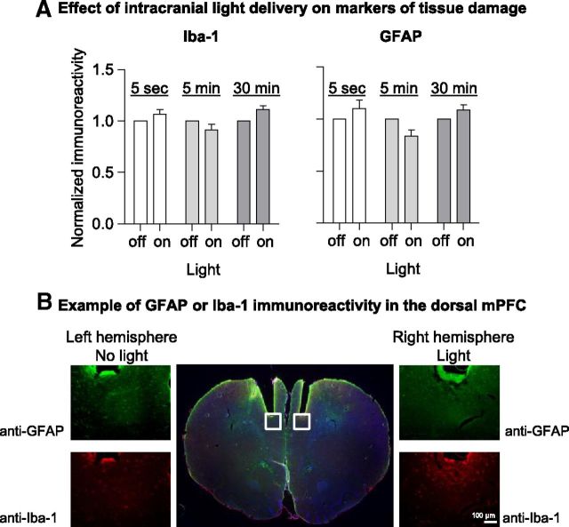
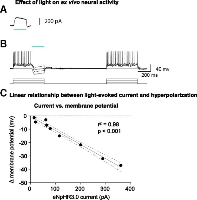
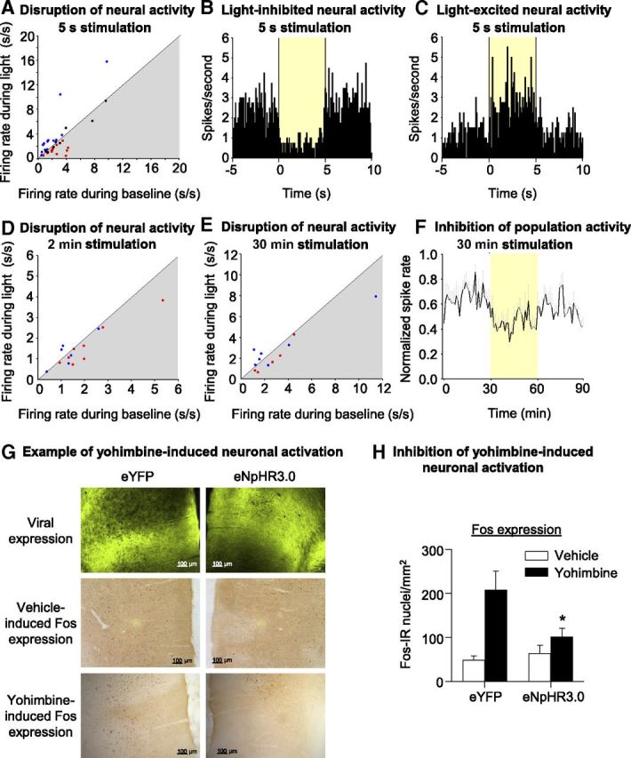
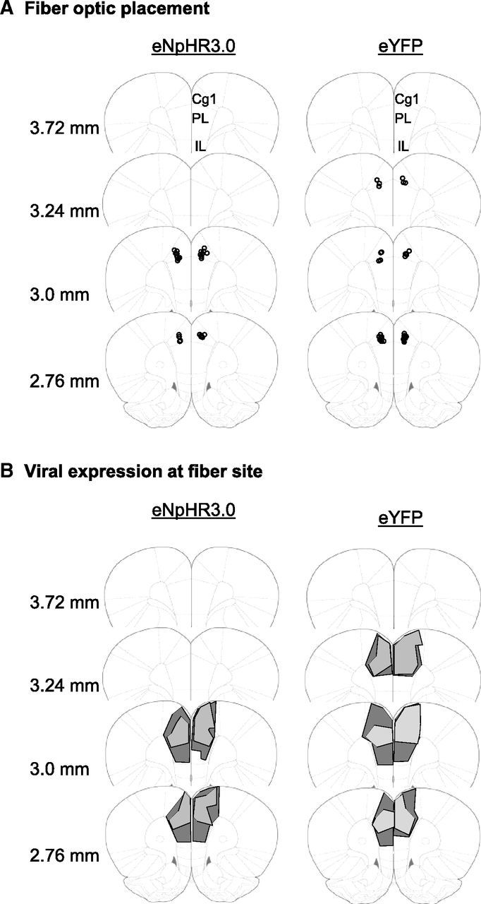
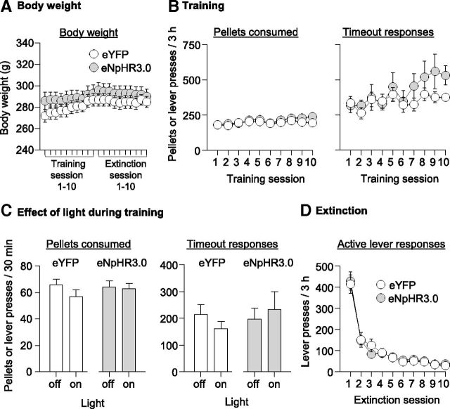
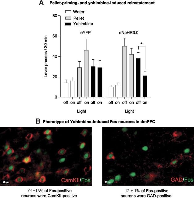
Relapse to maladaptive eating habits during dieting is often provoked by stress. Recently, we identified a role of dorsal medial prefrontal cortex (mPFC) neurons in stress-induced reinstatement of palatable food seeking in male rats. It is unknown whether endogenous neural activity in dorsal mPFC drives stress-induced reinstatement in female rats. Here, we used an optogenetic approach, in which female rats received bilateral dorsal mPFC microinjections of viral constructs coding light-sensitive eNpHR3.0-eYFP or control eYFP protein and intracranial fiber optic implants. Rats were food restricted and trained to lever press for palatable food pellets. Subsequently, pellets were removed, and lever pressing was extinguished; then the effect of bilateral dorsal mPFC light delivery on reinstatement of food seeking was assessed after injections of the pharmacological stressor yohimbine (an α-2 andrenoceptor antagonist) or pellet priming, a manipulation known to provoke food seeking in hungry rats. Dorsal mPFC light delivery attenuated yohimbine-induced reinstatement of food seeking in eNpHR3.0-injected but not eYFP-injected rats. This optical manipulation had no effect on pellet-priming-induced reinstatement or ongoing food-reinforced responding. Dorsal mPFC light delivery attenuated yohimbine-induced Fos immunoreactivity and disrupted neural activity during in vivo electrophysiological recording in awake rats. Optical stimulation caused significant outward currents and blocked electrically evoked action potentials in eNpHR3.0-injected but not eYFP-injected mPFC hemispheres. Light delivery alone caused no significant inflammatory response in mPFC. These findings indicate that intracranial light delivery in eNpHR3.0 rats disrupts endogenous dorsal mPFC neural activity that plays a role in stress-induced relapse to food seeking in female rats.


Figures

Figure 1.
Figure 1.
Intracranial light delivery and viral expression did not alter GFAP or Iba-1 immunoreactivity in the dorsal mPFC. A, Densitometric analysis of Iba-1 and GFAP immunoreactivity in the dorsal mPFC. All rats received 30 min of intracranial light delivery (593.5 nm, 20 mW), either as on/off cycles (5 s and 5 min) or as constant light (30 min). Immunoreactivity in the light-exposed hemisphere was compared with the control hemisphere (normalized to 1) for each rat. B, Representative immunohistochemical staining of astrocytes (GFAP) and microglia (Iba-1) in light-exposed (right) and control (left) hemispheres. 10× images correspond to the boxed areas of the central 1× image. Scale bar, 100 μm.
Figure 2.
Figure 2.
eNpHR3.0 activation decreased mPFC neuronal firing ex vivo. A, A 200 ms pulse of 532-nm light elicits robust outward current. B, Left, Action potentials elicited by increasing current steps (500 ms, +200 pA steps) are suppressed by eNpHR3.0 activation (200 ms light pulse). Right, Injected currents reliably evoke spikes in the absence of eNpHR3.0 activation. Green bar denotes time of light activation. C, A linear relationship is observed between eNpHR3.0-mediated currents and membrane hyperpolarization. Data are from 10 neurons from four rats.
Figure 3.
Figure 3.
Light delivery in vivo decreased dorsal mPFC neuronal activity in awake rats and also decreased yohimbine-induced neuronal activation. A, Scatter plot of all 35 neurons comparing activity during 5 s light-on versus no-light baseline period (baseline: mean of 5 s pre-light epoch and 5 s post-light epoch) for each neuron. Red dots represent neurons that were defined as light inhibited (fire significantly less during 5 s light-delivery epoch vs baseline). Blue dots represent neurons that were defined as light excited (fire significantly more during 5 s light-delivery epoch vs baseline). B, In vivo single-unit example of neural activity in a light-inhibited neuron in dorsal mPFC from eNpHR3.0-expressing rat. Perievent histogram showing average neural activity for a light-inhibited single unit across 40 trials of 5 s 593.5 nm, 4–8 mW intracranial light delivery. Activity (100 ms time bins) is aligned on light delivery, and the yellow box denotes duration of optical stimulation. C, In vivo single-unit example of neural activity in a light-excited neuron in dorsal mPFC from eNpHR3.0-expressing rat. Perievent histogram showing average neural activity for a light-inhibited single unit across 40 trials of 5 s 593.5 nm, 4–8 mW intracranial light delivery. Activity (100 ms time bins) is aligned on light delivery, and the yellow box denotes duration of optical stimulation. D, Scatter plot comparing activity during 2 min light-on versus 2 min no-light baseline period. Red and blue dots are the light-inhibited and light-excited neurons, respectively, as defined by their change in activity to the 5 s light stimulation parameter. Neurons that fall in the gray shaded area fire less during light delivery. E, Scatter plot comparing activity during 30 min light-on versus 30 min no-light baseline period. Red and blue dots are the light-inhibited and light-excited neurons, respectively, as defined by their change in activity to the 5 s light stimulation parameter. Neurons that fall in the gray shaded area fire less during light delivery. F, Mean ± SEM neural activity for a subpopulation of light-inhibited neurons (n = 5 neurons) during 30 min 593.5 nm, 4–8 mW intracranial light delivery. Activity (1 min time bins) is normalized to the maximum spike rate for each cell and is aligned on light delivery (yellow box denotes duration of optical stimulation). G, Example demonstrating the effect of bilateral intracranial light delivery on yohimbine-induced Fos expression. Example of sections demonstrating water-induced (middle row) and yohimbine-induced (bottom row) Fos expression in eYFP (left column) and eNpHR3.0 (right column) transfected hemispheres. Scale bars, 100 μm. H, Effect of light delivery on water- and yohimbine (2 mg/kg)-induced Fos expression in dorsal mPFC (n = 8 water; n = 9 yohimbine rats). Mean ± SEM counts of Fos-immunoreactive nuclei/mm2 in eYFP- and eNpHR3.0-expressing hemispheres of dorsal mPFC exposed to 90 min of 4–8 mW 593.5 laser light. Different from eYFP, *p < 0.05.
Figure 4.
Figure 4.
Histological confirmation of fiber optic placement and viral expression in dorsal mPFC in experiments 5 and 6. A, Circles represent the tip of the fiber optics, which are aimed ∼0.5 mm dorsal to the site of viral injection. B, Extent of viral expression at the site of fiber optic tip. Representative viral transduction in dorsal mPFC for the largest (dark gray) and smallest (light gray) area for eNpHR3.0–eYFP and eYFP expression at the site of the fiber optic tip. Note that this figure does not represent total viral spread but representative expression at the site of the fiber optic tip, in which presumably the light exerts the most potent effect on neural activity. Cg1, Cingulate cortex area 1; IL, infralimbic area; PL, prelimbic area.
Figure 5.
Figure 5.
Viral expression has no effect on body weight, acquisition of ongoing food self-administration, or extinction of food-reinforced responding, and intracranial light delivery had no effect on ongoing food self-administration. A, Body weight: mean ± SEM body weights during the training and extinction phases. B, Acquisition of food self-administration: mean ± SEM number of food pellets earned and timeout responses on the active lever (total lever presses minus pellets earned) during the training sessions. Rats were trained on a FR-1 20 s timeout reinforcement schedule; n = 17 eYFP and n = 15 eNpHR3.0. C, Effect of dorsal mPFC light delivery during ongoing food self-administration: mean ± SEM number of food pellets earned and timeout responses in the first 30 min of a training session when light was delivered and the subsequent training session for each rat when light was not delivered; n = 10 per virus condition. D, Extinction of food-reinforced responding: mean ± SEM number of responses on the previously active lever; n = 14 eYFP and n = 15 eNpHR3.0.
Figure 6.
Figure 6.
Intracranial light delivery decreased yohimbine-induced reinstatement but not pellet-priming-induced reinstatement of food seeking in eNpHR3.0 but not eYFP rats. A, Pellet priming- and yohimbine-induced reinstatement: mean ± SEM active lever presses in the first 30 min of a regular extinction session in which water injections were given before the session and the first 30 min of a test session in which either four noncontingent pellets were delivered at the beginning of the session or yohimbine injections (2 mg/kg, i.p) were given before testing. Data are shown for the control (intraperitoneal water injection) and the test sessions (pellet or yohimbine) with and without light delivery; n = 14 eYFP and n = 15 eNpHR3.0. *Different from the no-light (off) condition, p < 0.01. B, Phenotype of yohimbine-induced Fos neurons in dorsal mPFC: representative immunohistochemical staining of Fos+ and CaMKII+ (left) and Fos+ and GAD+ neurons (right). Scale bars, 20 μm.

References

    1. Adamantidis AR, Tsai HC, Boutrel B, Zhang F, Stuber GD, Budygin EA, Touriño C, Bonci A, Deisseroth K, de Lecea L. Optogenetic interrogation of dopaminergic modulation of the multiple phases of reward-seeking behavior. J Neurosci. 2011;31:10829–10835. - PMC - PubMed
    1. Anderson SM, Bari AA, Pierce RC. Administration of the D1-like dopamine receptor antagonist SCH-23390 into the medial nucleus accumbens shell attenuates cocaine priming-induced reinstatement of drug-seeking behavior in rats. Psychopharmacology. 2003;168:132–138. - PubMed
    1. Bossert JM, Stern AL, Theberge FR, Cifani C, Koya E, Hope BT, Shaham Y. Ventral medial prefrontal cortex neuronal ensembles mediate context-induced relapse to heroin. Nat Neurosci. 2011;14:420–422. - PMC - PubMed
    1. Bossert JM, Stern AL, Theberge FR, Marchant NJ, Wang HL, Morales M, Shaham Y. Role of projections from ventral medial prefrontal cortex to nucleus accumbens shell in context-induced reinstatement of heroin seeking. J Neurosci. 2012;32:4982–4991. - PMC - PubMed
    1. Boyden ES, Zhang F, Bamberg E, Nagel G, Deisseroth K. Millisecond-timescale, genetically targeted optical control of neural activity. Nat Neurosci. 2005;8:1263–1268. - PubMed

Publication types

LinkOut - more resources