

Specific inhibition of p25/Cdk5 activity by the Cdk5 inhibitory peptide reduces neurodegeneration in vivo
- PMID: 23283346
- PMCID: PMC6618624
- DOI: 10.1523/JNEUROSCI.3593-12.2013
Specific inhibition of p25/Cdk5 activity by the Cdk5 inhibitory peptide reduces neurodegeneration in vivo
Abstract
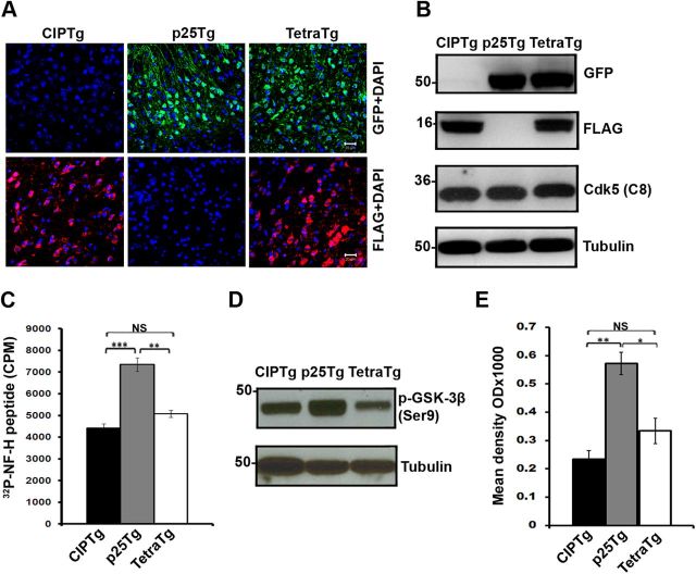
The aberrant hyperactivation of Cyclin-dependent kinase 5 (Cdk5), by the production of its truncated activator p25, results in the formation of hyperphosphorylated tau, neuroinflammation, amyloid deposition, and neuronal death in vitro and in vivo. Mechanistically, this occurs as a result of a neurotoxic insult that invokes the intracellular elevation of calcium to activate calpain, which cleaves the Cdk5 activator p35 into p25. It has been shown previously that the p25 transgenic mouse as a model to investigate the mechanistic implications of p25 production in the brain, which recapitulates deregulated Cdk5-mediated neuropathological changes, such as hyperphosphorylated tau and neuronal death. To date, strategies to inhibit Cdk5 activity have not been successful in targeting selectively aberrant activity without affecting normal Cdk5 activity. Here we show that the selective inhibition of p25/Cdk5 hyperactivation in vivo, through overexpression of the Cdk5 inhibitory peptide (CIP), rescues against the neurodegenerative pathologies caused by p25/Cdk5 hyperactivation without affecting normal neurodevelopment afforded by normal p35/Cdk5 activity. Tau and amyloid pathologies as well as neuroinflammation are significantly reduced in the CIP-p25 tetra transgenic mice, whereas brain atrophy and subsequent cognitive decline are reversed in these mice. The findings reported here represent an important breakthrough in elucidating approaches to selectively inhibit the p25/Cdk5 hyperactivation as a potential therapeutic target to reduce neurodegeneration.
Figures


Similar articles
-
AAV9-Mediated Cdk5 Inhibitory Peptide Reduces Hyperphosphorylated Tau and Inflammation and Ameliorates Behavioral Changes Caused by Overexpression of p25 in the Brain.J Alzheimers Dis. 2019;70(2):573-585. doi: 10.3233/JAD-190099. J Alzheimers Dis. 2019. PMID: 31256130
-
Involvement of aberrant cyclin-dependent kinase 5/p25 activity in experimental traumatic brain injury.J Neurochem. 2016 Jul;138(2):317-27. doi: 10.1111/jnc.13620. Epub 2016 May 25. J Neurochem. 2016. PMID: 26998748 Free PMC article.
-
Targeting Cdk5 activity in neuronal degeneration and regeneration.Cell Mol Neurobiol. 2009 Dec;29(8):1073-80. doi: 10.1007/s10571-009-9410-6. Cell Mol Neurobiol. 2009. PMID: 19455415 Free PMC article.
-
Peptides derived from Cdk5 activator p35, specifically inhibit deregulated activity of Cdk5.Biotechnol J. 2007 Aug;2(8):978-87. doi: 10.1002/biot.200700057. Biotechnol J. 2007. PMID: 17526058 Review.
-
Deregulated Cdk5 activity is involved in inducing Alzheimer's disease.Arch Med Res. 2012 Nov;43(8):655-62. doi: 10.1016/j.arcmed.2012.10.015. Epub 2012 Nov 7. Arch Med Res. 2012. PMID: 23142263 Free PMC article. Review.
Cited by
-
Cyclin-dependent Kinase 5 and Neurodegenerative Diseases.Mol Neurobiol. 2024 Oct;61(10):7287-7302. doi: 10.1007/s12035-024-04047-1. Epub 2024 Feb 20. Mol Neurobiol. 2024. PMID: 38378992 Review.
-
The Cyclin-Dependent Kinase 5 Inhibitor Peptide Inhibits Herpes Simplex Virus Type 1 Replication.Sci Rep. 2019 Feb 4;9(1):1260. doi: 10.1038/s41598-018-37989-3. Sci Rep. 2019. PMID: 30718749 Free PMC article.
-
Cdk5 links with DNA damage response and cancer.Mol Cancer. 2017 Mar 14;16(1):60. doi: 10.1186/s12943-017-0611-1. Mol Cancer. 2017. PMID: 28288624 Free PMC article. Review.
-
Biological functions of CDK5 and potential CDK5 targeted clinical treatments.Oncotarget. 2017 Mar 7;8(10):17373-17382. doi: 10.18632/oncotarget.14538. Oncotarget. 2017. PMID: 28077789 Free PMC article. Review.
-
When Good Kinases Go Rogue: GSK3, p38 MAPK and CDKs as Therapeutic Targets for Alzheimer's and Huntington's Disease.Int J Mol Sci. 2021 May 31;22(11):5911. doi: 10.3390/ijms22115911. Int J Mol Sci. 2021. PMID: 34072862 Free PMC article. Review.
References
-
- Ahlijanian MK, Barrezueta NX, Williams RD, Jakowski A, Kowsz KP, McCarthy S, Coskran T, Carlo A, Seymour PA, Burkhardt JE, Nelson RB, McNeish JD. Hyperphosphorylated tau and neurofilament and cytoskeletal disruptions in mice overexpressing human p25, an activator of cdk5. Proc Natl Acad Sci U S A. 2000;97:2910–2915. - PMC - PubMed
-
- Amin ND, Albers W, Pant HC. Cyclin-dependent kinase 5 (cdk5) activation requires interaction with three domains of p35. J Neurosci Res. 2002;67:354–362. - PubMed
-
- Aoki I, Wu YJ, Silva AC, Lynch RM, Koretsky AP. In vivo detection of neuroarchitecture in the rodent brain using manganese-enhanced MRI. Neuroimage. 2004;22:1046–1059. - PubMed
-
- Bian F, Nath R, Sobocinski G, Booher RN, Lipinski WJ, Callahan MJ, Pack A, Wang KK, Walker LC. Axonopathy, tau abnormalities, and dyskinesia, but no neurofibrillary tangles in p25-transgenic mice. J Comp Neurol. 2002;446:257–266. - PubMed
-
- Chuang KH, Koretsky A. Improved neuronal tract tracing using manganese enhanced magnetic resonance imaging with fast T(1) mapping. Magn Reson Med. 2006;55:604–611. - PubMed
Publication types
MeSH terms
Substances
LinkOut - more resources
Full Text Sources
Other Literature Sources
Molecular Biology Databases
Research Materials