

The functional architecture of S1 during touch observation described with 7 T fMRI
- PMID: 23283478
- PMCID: PMC3889700
- DOI: 10.1007/s00429-012-0489-z
The functional architecture of S1 during touch observation described with 7 T fMRI
Abstract
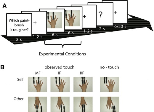
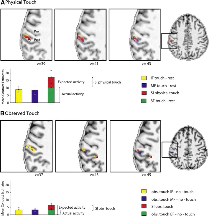
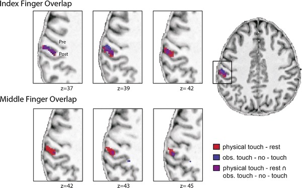
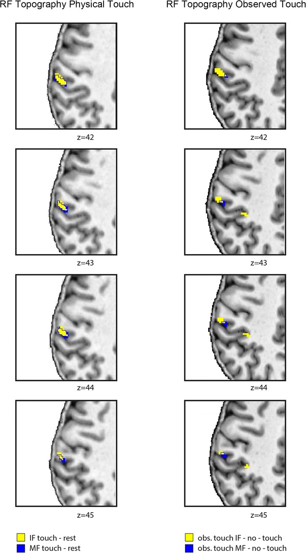
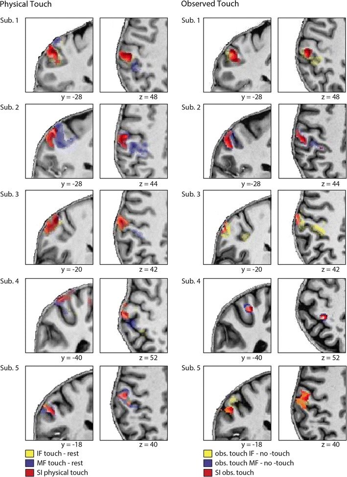
Recent studies indicate that the primary somatosensory cortex (S1) is active not only when touch is physically perceived but also when it is merely observed to be experienced by another person. This social responsivity of S1 has important implications for our understanding of S1 functioning. However, S1 activity during touch observation has not been characterized in great detail to date. We focused on two features of the S1 functional architecture during touch observation, namely the topographical arrangement of index and middle finger receptive fields (RFs), and their dynamic shrinkage during concurrent activation. Both features have important implications for human behavior. We conducted two fMRI studies at 7 T, one where touch was physically perceived, and one where touch was observed. In the two experiments, participants either had their index finger and/or middle finger stimulated using paintbrushes, or just observed similar touch events on video. Our data show that observing and physically experiencing touch elicits overlapping activity changes in S1. In addition, observing touch to the index finger or the middle finger alone evoked topographically arranged activation foci in S1. Importantly, when co-activated, the index and middle finger RFs not only shrank during physical touch perception, but also during touch observation. Our data, therefore, indicate a similarity between the functional architecture of S1 during touch observation and physical touch perception with respect to single-digit topography and RF shrinkage. These results may allow the tentative conclusion that even primary somatosensory experiences, such as physical touch perception, can be shared amongst individuals.
Figures
Similar articles
-
Judging roughness by sight--a 7-Tesla fMRI study on responsivity of the primary somatosensory cortex during observed touch of self and others.Hum Brain Mapp. 2013 Aug;34(8):1882-95. doi: 10.1002/hbm.22031. Epub 2012 Mar 16. Hum Brain Mapp. 2013. PMID: 22422484 Free PMC article.
-
Seeing is not feeling: posterior parietal but not somatosensory cortex engagement during touch observation.J Neurosci. 2015 Jan 28;35(4):1468-80. doi: 10.1523/JNEUROSCI.3621-14.2015. J Neurosci. 2015. PMID: 25632124 Free PMC article.
-
Understanding others' feelings: the role of the right primary somatosensory cortex in encoding the affective valence of others' touch.J Neurosci. 2013 Feb 27;33(9):4201-5. doi: 10.1523/JNEUROSCI.4498-12.2013. J Neurosci. 2013. PMID: 23447627 Free PMC article.
-
Observing touch activates human primary somatosensory cortex.Eur J Neurosci. 2010 May;31(10):1836-43. doi: 10.1111/j.1460-9568.2010.07192.x. Eur J Neurosci. 2010. PMID: 20584188
-
Tactile acuity of fingertips and hand representation size in human Area 3b and Area 1 of the primary somatosensory cortex.Neuroimage. 2021 May 15;232:117912. doi: 10.1016/j.neuroimage.2021.117912. Epub 2021 Feb 27. Neuroimage. 2021. PMID: 33652142
Cited by
-
How Visual Body Perception Influences Somatosensory Plasticity.Neural Plast. 2018 Mar 11;2018:7909684. doi: 10.1155/2018/7909684. eCollection 2018. Neural Plast. 2018. PMID: 29713338 Free PMC article. Review.
-
Lower visual field preference for the visuomotor control of limb movements in the human dorsomedial parietal cortex.Brain Struct Funct. 2021 Dec;226(9):2989-3005. doi: 10.1007/s00429-021-02254-3. Epub 2021 Mar 18. Brain Struct Funct. 2021. PMID: 33738579 Free PMC article.
-
Body maps in the infant brain.Trends Cogn Sci. 2015 Sep;19(9):499-505. doi: 10.1016/j.tics.2015.06.012. Epub 2015 Jul 28. Trends Cogn Sci. 2015. PMID: 26231760 Free PMC article. Review.
-
Shared neural representations of tactile roughness intensities by somatosensation and touch observation using an associative learning method.Sci Rep. 2019 Jan 11;9(1):77. doi: 10.1038/s41598-018-37378-w. Sci Rep. 2019. PMID: 30635598 Free PMC article.
-
Seeing Touches Early in Life.PLoS One. 2015 Sep 14;10(9):e0134549. doi: 10.1371/journal.pone.0134549. eCollection 2015. PLoS One. 2015. PMID: 26366563 Free PMC article.
References
Publication types
MeSH terms
Substances
LinkOut - more resources
Full Text Sources
Other Literature Sources
Medical
