

Shared cell surface marker expression in mesenchymal stem cells and adult sarcomas
- PMID: 23283492
- PMCID: PMC3659743
- DOI: 10.5966/sctm.2012-0055
Shared cell surface marker expression in mesenchymal stem cells and adult sarcomas
Abstract
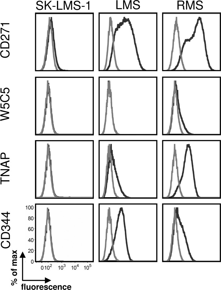
Advanced adult soft-tissue sarcomas (STSs) are rare tumors with a dismal prognosis and limited systemic treatment options. STSs may originate from mesenchymal stem cells (MSCs); the latter have mainly been isolated from adult bone marrow as plastic-adherent cells with differentiation capacity into mesenchymal tissues. Recently, a panel of antibodies has been established that allows for the prospective isolation of primary MSCs with high selectivity. Similar to cancer stem cells in other malignancies, sarcoma stem cells may bear immunophenotypic similarity with the corresponding precursor, that is, MSCs. We therefore set out to establish the expression pattern of MSC markers in sarcoma cell lines and primary tumor samples by flow cytometry. In addition, fibroblasts from different sources were examined. The results document a significant amount of MSC markers shared by sarcoma cells. The expression pattern includes uniformly expressed markers, as well as MSC markers that only stained subpopulations of sarcoma cells. Expression of W5C5, W8B2 (tissue nonspecific alkaline phosphatase [TNAP]), CD344 (frizzled-4), and CD271 marked subpopulations displaying increased proliferation potential. Moreover, CD271+ cells displayed in vitro doxorubicin resistance and an increased capacity to form spheres under serum-free conditions. Interestingly, another set of antigens, including the bona fide progenitor cell markers CD117 and CD133, were not expressed. Comparative expression patterns of novel MSC markers in sarcoma cells, as well as fibroblasts and MSCs, are presented. Our data suggest a hierarchical cytoarchitecture of the most common adult type sarcomas and introduce W5C5, TNAP, CD344, and CD271 as potential sarcoma progenitor cell markers.
Figures


Similar articles
-
Chondrogenic potential of subpopulations of cells expressing mesenchymal stem cell markers derived from human synovial membranes.J Cell Biochem. 2010 Nov 1;111(4):834-45. doi: 10.1002/jcb.22768. J Cell Biochem. 2010. PMID: 20665538
-
Characterization of different subpopulations from bone marrow-derived mesenchymal stromal cells by alkaline phosphatase expression.Stem Cells Dev. 2012 Nov 1;21(16):2958-68. doi: 10.1089/scd.2011.0349. Epub 2012 Jul 18. Stem Cells Dev. 2012. PMID: 22702738 Free PMC article.
-
The CD133+ subpopulation of the SW982 human synovial sarcoma cell line exhibits cancer stem-like characteristics.Int J Oncol. 2013 Apr;42(4):1399-407. doi: 10.3892/ijo.2013.1826. Epub 2013 Feb 15. Int J Oncol. 2013. PMID: 23416969
-
Modeling sarcomagenesis using multipotent mesenchymal stem cells.Cell Res. 2012 Jan;22(1):62-77. doi: 10.1038/cr.2011.157. Epub 2011 Sep 20. Cell Res. 2012. PMID: 21931359 Free PMC article. Review.
-
Epigenetic remodeling of chromatin architecture: exploring tumor differentiation therapies in mesenchymal stem cells and sarcomas.Curr Stem Cell Res Ther. 2010 Mar;5(1):63-73. doi: 10.2174/157488810790442859. Curr Stem Cell Res Ther. 2010. PMID: 19807660 Free PMC article. Review.
Cited by
-
Mesenchymal stromal cells in cancer: a review of their immunomodulatory functions and dual effects on tumor progression.J Pathol. 2020 Apr;250(5):555-572. doi: 10.1002/path.5357. Epub 2019 Dec 18. J Pathol. 2020. PMID: 31608444 Free PMC article. Review.
-
Activity of cytokine-induced killer cells against bone and soft tissue sarcoma.Oncoimmunology. 2014 Mar 17;3:e28269. doi: 10.4161/onci.28269. eCollection 2014. Oncoimmunology. 2014. PMID: 25050197 Free PMC article.
-
Platelet-derived growth factor receptor-α and -β promote cancer stem cell phenotypes in sarcomas.Oncogenesis. 2018 Jun 19;7(6):47. doi: 10.1038/s41389-018-0059-1. Oncogenesis. 2018. Retraction in: Oncogenesis. 2024 May 29;13(1):19. doi: 10.1038/s41389-024-00520-7. PMID: 29915281 Free PMC article. Retracted.
-
Quality Control of Stem Cell-Based Cultured Meat According to Specific Differentiation Abilities.Cells. 2024 Jan 11;13(2):135. doi: 10.3390/cells13020135. Cells. 2024. PMID: 38247826 Free PMC article.
-
Cancer Stem Cells in Sarcomas: In Vitro Isolation and Role as Prognostic Markers: A Systematic Review.Cancers (Basel). 2023 Apr 25;15(9):2449. doi: 10.3390/cancers15092449. Cancers (Basel). 2023. PMID: 37173919 Free PMC article. Review.
References
-
- Kopp HG, Patel S, Brucher B, et al. Potential combination chemotherapy approaches for advanced adult-type soft-tissue sarcoma. Am J Clin Dermatol. 2008;9:207–217. - PubMed
-
- Jemal A, Siegel R, Ward E, et al. Cancer statistics, 2009. CA Cancer J Clin. 2009;59:225–249. - PubMed
-
- Fletcher CDM, Unni KK, Mertens F, editors. WHO Classification of Tumours of Soft Tissue and Bone. Lyon, France: IARC Press; 2002. Pathology and Genetics of Tumours of Soft Tissue and Bone.
-
- Katenkamp D, Kosmehl H. Heterogeneity in malignant soft tissue tumors. Curr Top Pathol. 1995;89:123–151. - PubMed
-
- Katenkamp D. Cellular heterogeneity: Explanation for changing of tumor phenotype and biologic behavior in soft tissue sarcomas. Pathol Res Pract. 1988;183:698–705. - PubMed
Publication types
MeSH terms
Substances
LinkOut - more resources
Full Text Sources
Medical
Research Materials
