. 2012 Dec;1(12):855-65.
doi: 10.5966/sctm.2012-0093. Epub 2012 Nov 29.

A practical and efficient cellular substrate for the generation of induced pluripotent stem cells from adults: blood-derived endothelial progenitor cells

Affiliations

A practical and efficient cellular substrate for the generation of induced pluripotent stem cells from adults: blood-derived endothelial progenitor cells

Imbisaat Geti et al. Stem Cells Transl Med. 2012 Dec.

Abstract

Induced pluripotent stem cells (iPSCs) have the potential to generate patient-specific tissues for disease modeling and regenerative medicine applications. However, before iPSC technology can progress to the translational phase, several obstacles must be overcome. These include uncertainty regarding the ideal somatic cell type for reprogramming, the low kinetics and efficiency of reprogramming, and karyotype discrepancies between iPSCs and their somatic precursors. Here we describe the use of late-outgrowth endothelial progenitor cells (L-EPCs), which possess several favorable characteristics, as a cellular substrate for the generation of iPSCs. We have developed a protocol that allows the reliable isolation of L-EPCs from peripheral blood mononuclear cell preparations, including frozen samples. As a proof-of-principle for clinical applications we generated EPC-iPSCs from both healthy individuals and patients with heritable and idiopathic forms of pulmonary arterial hypertension. L-EPCs grew clonally; were highly proliferative, passageable, and bankable; and displayed higher reprogramming kinetics and efficiencies compared with dermal fibroblasts. Unlike fibroblasts, the high efficiency of L-EPC reprogramming allowed for the reliable generation of iPSCs in a 96-well format, which is compatible with high-throughput platforms. Array comparative genome hybridization analysis of L-EPCs versus donor-matched circulating monocytes demonstrated that L-EPCs have normal karyotypes compared with their subject's reference genome. In addition, >80% of EPC-iPSC lines tested did not acquire any copy number variations during reprogramming compared with their parent L-EPC line. This work identifies L-EPCs as a practical and efficient cellular substrate for iPSC generation, with the potential to address many of the factors currently limiting the translation of this technology.


Figures
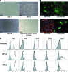
Figure 1.
Figure 1.
Generation and characterization of L-EPCs. (A): Generation of L-EPCs. Late-outgrowth endothelial progenitor cells emerged from within cultures of early endothelial progenitor cells. Early-EPCs (E-EPCs) formed adherent cultures with a monocyte-derived macrophage type morphology (day 7) and predominated in the culture flask up to around day 15; however, L-EPCs emerged by day 10, forming highly proliferative colonies (day 13) of cells resembling endothelial cells, and became the predominant cell type in the flask exhibiting the cobblestone morphology shared with endothelial cell cultures (day 18). L-EPCs were able to form endothelial cell-like networks in vitro. (B): Characterization of established L-EPCs. Immunostaining of L-EPCs at passage 4 revealed expression of endothelial cell-specific markers vWF (green; DAPI, blue), CD146 (green; DAPI, blue), and CD31 (red; DAPI, blue) and also the progenitor cell marker CD34 (green; DAPI, blue). (C): Flow cytometric analysis demonstrated that L-EPCs (passage 4) expressed surface marker expression similar to that of pulmonary artery endothelial cells when compared with freshly isolated monocytes (which predominated in the E-EPC population). Abbreviations: DAPI, 4′,6-diamidino-2-phenylindole; L-EPC, late-outgrowth endothelial progenitor cell; KDR, kinase insert domain receptor; PAEC, pulmonary artery endothelial cell; vWF, von Willebrand factor.
Figure 2.
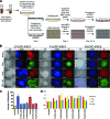
Figure 2.
Summary of methodology and basic characterization of EPC-iPSCs. (A): Overview of EPC-iPSC derivation. iPSC derivation in standard 10-cm dishes and 96-well high-throughput method. Mononuclear cells are isolated from nonmobilized peripheral blood. These cells are transduced with retrovirus expressing Oct4, Sox2, c-Myc, and KLF4 and either added to MEFs or MEFs are added to infected late-outgrowth endothelial progenitor cell (L-EPC) cultures. Colonies of iPSCs emerge and are either picked or stained. (B): Pluripotency marker expression in three exemplar lines of iPSCs generated from EPCs. Panels in the first group of columns (C7-EPC-iPSC1) are subline 1 of iPSCs derived from L-EPC line C7-EPC (from normal control). Panels in the middle group of columns (P1-EPC-iPSC1) are subline 1 of iPSCs derived from L-EPC line P1-EPC (from a patient with pulmonary arterial hypertension [PAH] harboring a mutation in bone morphogenetic protein type II receptor (BMPR2). Panels in the third group of columns (P3-EPC-iPSC2) are subline 2 of iPSCs derived from L-EPC line P3-EPC (from a patient with PAH without an identifiable mutation in BMPR2). All iPSC lines expressed the markers NANOG, OCT4, SOX2, and TRA-1-60, which is consistent with a pluripotent state. (C): Relative levels of DNA methylation on the Oct4 promoter of EPC-iPSCs generated from L-EPC compared with H9 hESCs. L-EPC lines C7-EPC and P3-EPC3 were analyzed along with their derivative iPSC sublines 1, 2, and 9 and 2 and 3, respectively. (D): Viral transgene insertion rates of EPC-iPSCs generated from L-EPC lines C7-EPC (see Results section and supplemental online Table 6) and P3-EPC (see Results section and supplemental online Table 6). The number of viral integration was obtained by quantitative polymerase chain reaction genotyping analyses quantifying the number of copies of each gene relative to the endogenous levels of each gene in H9 hESCs. Thus, the H9 control has two copies represented as one on the y-axis. Abbreviations: BF, bright field; C, control; DAPI, 4′,6-diamidino-2-phenylindole; EPC, endothelial progenitor cell; FCS, fetal calf serum; hESC, human embryonic stem cell; IF, immunofluorescence; iPSC, induced pluripotent stem cell; MEF, mouse embryonic fibroblast; P, patient.
Figure 3.
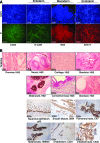
Figure 3.
Differentiation of endothelial progenitor cell (EPC)-induced pluripotent stem cells (iPSCs). (A): A single iPSC exemplar line (C7-EPC-iPSC1). EPC-iPSCs could be differentiated using chemically defined differentiation protocols in vitro into derivatives of the three germ layers, ectoderm (N-CAM), mesoderm (BRA), and endoderm (SOX17), and tissues expressing posterior mesoderm/extraembryonic tissues (CDX2). (B): Two examplar lines of EPC-iPSCs (C7-EPC-iPSC1 and P1-EPC-iPSC3). In in vivo teratoma analyses EPC-iPSCs also differentiated into derivatives of the three germ layers identifiable by H&E staining (bottom two rows) or using specific antibodies (ectodermal derivatives, P63, HMB45; mesodermal derivatives, SMA, CD31; endoderm, CK7, CK20). Abbreviations: BRA, brachyury; CK, cytokeratin; DAPI, 4′,6-diamidino-2-phenylindole; H&E, hematoxylin and eosin; IF, immunofluorescence; N-CAM, neural cell adhesion molecule; SMA, smooth muscle actin.
Figure 4.
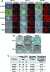
Figure 4.
Kinetics of iPSC generation from late-outgrowth endothelial progenitor cells (L-EPCs). (A): NANOG-expressing colonies were detected from day 10 of reprogramming of L-EPCs and day 15 of fibroblasts (day 10 NANOG expression panels are shown at twice the magnification of all other panels). (B): Examplar comparison of iPSC derivation efficiency between C7-EPC and Fibro1. Each well contained 33,333 infected cells. iPSC colonies stained with alkaline phosphatase (AP) positive at different time points. Scale bars = 8.75 mm. (C): Frequencies of AP-positive colonies derived from three L-EPC lines and two fibroblast lines. Experimental design was the same as described for (B). Abbreviations: C, control; EPC, endothelial progenitor cell; Fibro, fibroblast; iPSC, induced pluripotent stem cell; P, patient.
Figure 5.
Figure 5.
Ninety-six-well format reprogramming of late-outgrowth endothelial progenitor cells (L-EPCs). (A): 4,000 L-EPCs or fibroblasts were added to one well of a 96-well dish. This was replicated 11 times, so that every well in a row had 4,000 cells of the same line. These were infected with the reprogramming viruses, and on day 5 mouse embryonic fibroblasts were added to the wells. Alkaline phosphatase (AP) staining was carried out on day 15, and induced pluripotent stem cell colonies appeared blue/purple. (B): Frequencies of AP-positive iPSC colonies per well. Abbreviations: C, control; EPC, endothelial progenitor cell; P, patient.

References

    1. Takahashi K, Yamanaka S. Induction of pluripotent stem cells from mouse embryonic and adult fibroblast cultures by defined factors. Cell. 2006;126:663–676. - PubMed
    1. Takahashi K, Tanabe K, Ohnuki M, et al. Induction of pluripotent stem cells from adult human fibroblasts by defined factors. Cell. 2007;131:861–872. - PubMed
    1. Hussein SM, Batada NN, Vuoristo S, et al. Copy number variation and selection during reprogramming to pluripotency. Nature. 2011;471:58–62. - PubMed
    1. Martins-Taylor K, Nisler BS, Taapken SM, et al. Recurrent copy number variations in human induced pluripotent stem cells. Nat Biotechnol. 2011;29:488–491. - PubMed
    1. Yusa K, Rashid ST, Strick-Marchand H, et al. Targeted gene correction of α1-antitrypsin deficiency in induced pluripotent stem cells. Nature. 2011;478:391–394. - PMC - PubMed

Publication types

MeSH terms