. 2012 Dec;1(12):886-97.
doi: 10.5966/sctm.2012-0086. Epub 2012 Nov 29.

Efficient lentiviral transduction of human mesenchymal stem cells that preserves proliferation and differentiation capabilities

Affiliations

Efficient lentiviral transduction of human mesenchymal stem cells that preserves proliferation and differentiation capabilities

Paul Lin et al. Stem Cells Transl Med. 2012 Dec.

Abstract

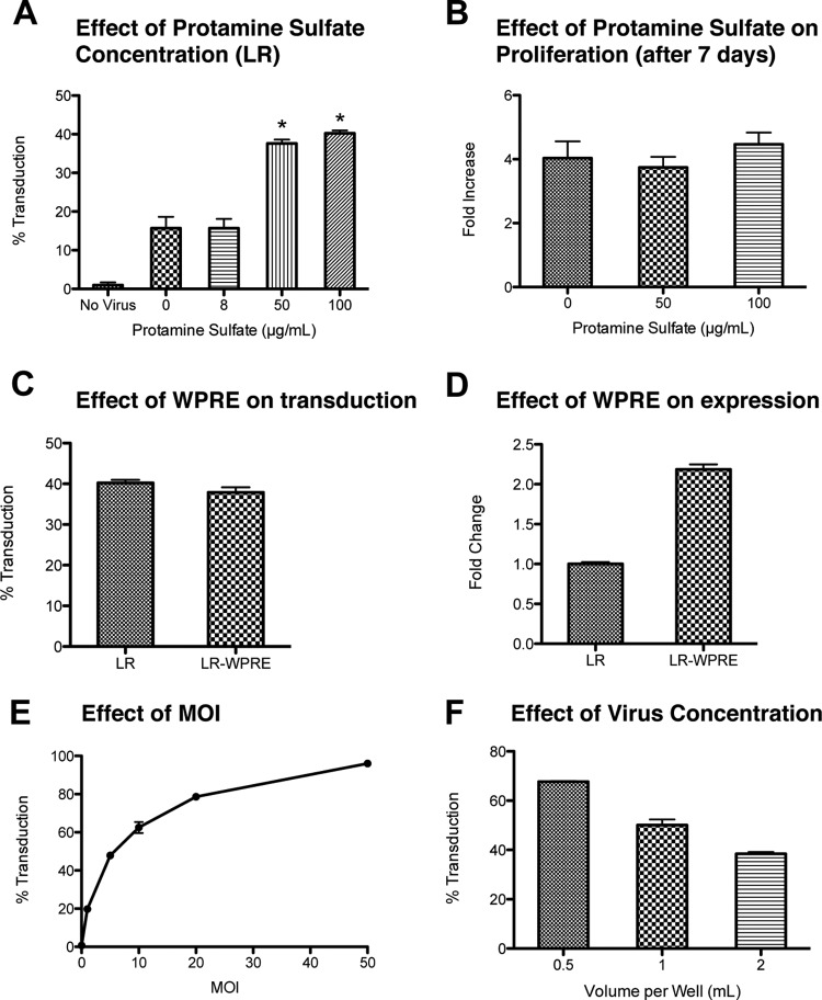
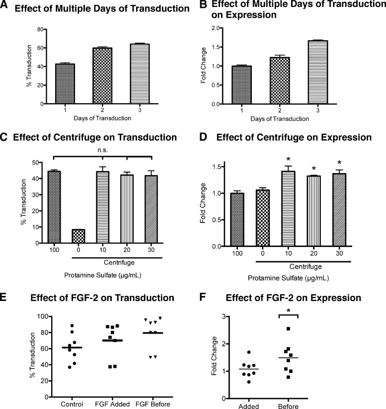
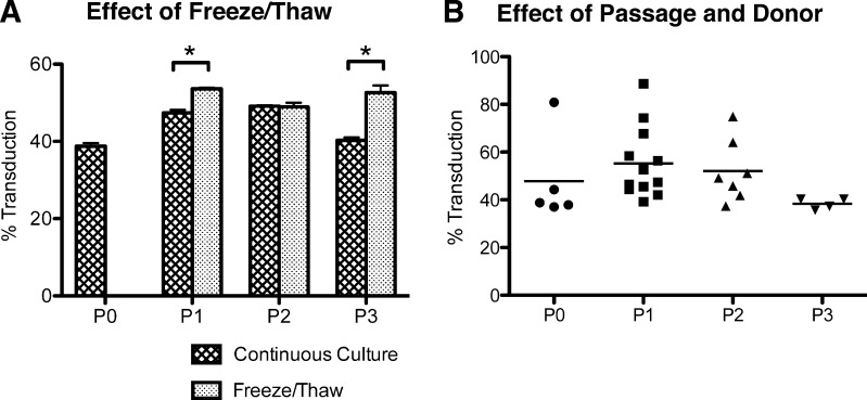
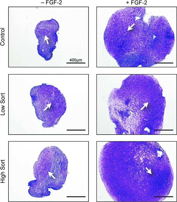
Long-term lentiviral transduction of human mesenchymal stem cells (hMSCs) greatly enhances the usefulness of these cells. However, such transduction currently requires the use of polybrene, which severely inhibits hMSC proliferation. In contrast, protamine sulfate at 100 μg/ml doubled transduction efficiencies without affecting proliferation or differentiation potential. Expression levels improved 2.2-fold with the addition of a woodchuck hepatitis post-transcriptional regulatory element. Further improvements in transduction efficiencies could be obtained by a modest increase in viral concentrations through increased viral titers or decreased transduction volumes without changing multiplicity of infection, by transducing over multiple days, or by culturing the cells in fibroblast growth factor-2. Centrifugation improved expression but had no effect on efficiency. Transgene expression was stable over 6 weeks in vitro and in vivo. Donor-to-donor and intradonor variability were observed in primary passage through passage 2 cultures, but not at passage 3. These results provide a better optimized approach for expanded use of hMSCs through genetic manipulation.


Figures

Figure 1.
Figure 1.
Effect of protamine sulfate, WPRE, and viral concentration. (A): Human mesenchymal stem cells (hMSCs) were incubated with different concentrations of protamine sulfate at a constant concentration of LR coding for a luciferase/monomeric red fluorescent fusion protein. The transduction efficiency was measured by the red fluorescent intensity. *, p < .001 vs. 0 and 8 μg/ml, Tukey's t test. MOI = 5. (B): The increase in cell numbers was measured after 1 week in culture for hMSCs cultured in 0 μg/ml protamine sulfate (control) and at different concentrations of protamine sulfate. There was no statistical difference between the groups. (C): Transduction efficiency of the lentivirus with WPRE (LR-WPRE) or without WPRE (LR). No statistical difference (t test). (D): Difference in relative fluorescence values between the gene construct with or without WPRE. p < .001, t test. (E): Transduction efficiency of hMSCs incubated at different MOI but the same transduction volume (1 ml per well of a six-well plate) and protamine sulfate concentration (100 μg/ml). (F): Transduction efficiency at different transduction volumes in a six-well plate, but the same MOI (MOI = 5) and protamine sulfate concentration (100 μg/ml); p < .001 between all groups. All values are mean ± SD. Abbreviations: LR, lentivirus with luciferase and monomeric red fluorescent protein; MOI, multiplicity of infection; WPRE, woodchuck hepatitis post-transcriptional regulatory element.
Figure 2.
Figure 2.
Optimizing transduction efficiency and transgene expression. (A, B): The change in transduction efficiency (p < .05 between all groups) (A) and relative change in transgene expression (p < .01 between all groups) (B) of transducing over 2 to 3 days compared with a 1-day transduction. The total amount of virus was kept the same. For 1 day, MOI = 5; 2 days, MOI = 2.5; and 3 days, MOI = 1.67. Transduction volume (1 ml per well of a six-well plate) and protamine sulfate concentration (100 μg/ml) were held constant. Expression levels were compared with 1-day transduction. Values are mean ± SD. (C, D): The change in transduction efficiency (C) and relative change in transgene expression (D) of cells centrifuged with different concentrations of protamine sulfate compared with hMSCs transduced in static culture at 100 μg/ml protamine sulfate. At 10–30 μg/ml protamine sulfate, there was no statistical difference in transduction efficiency between static culture and centrifugation, whereas there was a statistical difference in transgene expression. *, p < .01 vs. static 100 μg/ml and centrifuge 0 μg/ml, Tukey's t test. Expression levels were compared with static culture. MOI = 5. Values are mean ± SD. (E, F): The change in transduction efficiency (p < .05, between all conditions, n = 8) (E) and transgene expression (*, p < .05, n = 8) (F) of hMSCs treated with FGF-2 prior to transduction (before) or at the time of transduction (added) compared with hMSCs cultured without FGF-2 (control). Expression levels were compared with control. All experiments involving FGF-2 were done with 100 μg/ml protamine sulfate and MOI = 5. Each data point is a mean of three replicates of one experiment. Abbreviation: FGF-2, fibroblast growth factor-2.
Figure 3.
Figure 3.
Consistency of MSC transduction. (A): The transduction efficiency of hMSCs frozen and thawed at different passages compared with those that were maintained continuously in culture. MOI = 5; protamine sulfate = 100 μg/ml. Values are mean ± SD. *, p < .001. (B): A graph of all the transductions from the different donors at different passages. n = 5, 12, 7, and 4 respectively for P0, P1, P2, and P3. MOI = 5; protamine sulfate = 100 μg/ml. No statistical significance was found between passages or within donors. Each data point is a mean of three replicates of one experiment. Abbreviation: P, passage.
Figure 4.
Figure 4.
Chondrogenic pellet assay. Human mesenchymal stem cells were transduced with or without prior FGF-2 treatment and then sorted into the top 30% highest expressing (high sort) and bottom 30% lowest expressing (low sort) cells and tested for their ability to form cartilage in pellet cultures compared with nontransduced cells. The white arrows point to the cartilage matrix. Scale bars = 400 μm. Abbreviation: FGF-2, fibroblast growth factor-2.
Figure 5.
Figure 5.
Adipogenic assay. Human mesenchymal stem cells were cultured in adipogenic medium for 21 days. The cultures were imaged for red fluorescence and merged with the immunofluorescence stain for adipophilin (green). Transduced cells show both red fluorescence and adipophilin staining. Scale bars = 50 μm. Abbreviation: FGF-2, fibroblast growth factor-2.
Figure 6.
Figure 6.
Ceramic cube assay. (A): Sorted human mesenchymal stem cells (hMSCs) were seeded into ceramic cubes and implanted subcutaneously. The transduced hMSCs continued to show strong bioluminescence after 6 weeks in vivo. An x-ray image shows the location of the ceramic cubes. Numbers indicate: hMSCs cultured without FGF-2, high sort (1) and low sort (4); hMSCs cultured with FGF-2, high sort (5) and low sort (7); nontransduced hMSCs (2, 3, 6). (B): The high sort cells were cultured for another seven passages, equivalent to 7 weeks, and their transduction efficiency was measured at each passage. The numbers in the bars indicate the passage numbers. Abbreviation: FGF-2, fibroblast growth factor-2.
Figure 7.
Figure 7.
Ceramic cube assay. After 6 weeks, the ceramic cubes were harvested and stained for histologic analysis. The black arrows point to the bone matrix found in all ceramics. FGF-2-treated cells showed more bone matrix overall. Scale bars = 200 μm. Abbreviation: FGF-2, fibroblast growth factor-2.

References

    1. Caplan AI. Mesenchymal stem cells. J Orthop Res. 1991;9:641–650. - PubMed
    1. Pittenger MF, Mackay AM, Beck SC, et al. Multilineage potential of adult human mesenchymal stem cells. Science. 1999;284:143–147. - PubMed
    1. Kadiyala S, Jaiswal N, Bruder SP. Culture-expanded, bone marrow-derived mesenchymal stem cells can regenerate a critical-sized segmental bone defect. Tissue Eng. 1997;3:173–185.
    1. Solchaga LA, Penick KJ, Welter JF. Chondrogenic differentiation of bone marrow-derived mesenchymal stem cells: Tips and tricks. Methods Mol Biol. 2011;698:253–278. - PMC - PubMed
    1. Bai L, Lennon DP, Eaton V, et al. Human bone marrow-derived mesenchymal stem cells induce Th2-polarized immune response and promote endogenous repair in animal models of multiple sclerosis. Glia. 2009;57:1192–1203. - PMC - PubMed

Publication types

MeSH terms