. 2012 Dec;1(12):921-6.
doi: 10.5966/sctm.2012-0026. Epub 2012 Nov 26.

Angiogenic dysfunction in bone marrow-derived early outgrowth cells from diabetic animals is attenuated by SIRT1 activation

Affiliations

Angiogenic dysfunction in bone marrow-derived early outgrowth cells from diabetic animals is attenuated by SIRT1 activation

Darren A Yuen et al. Stem Cells Transl Med. 2012 Dec.

Abstract

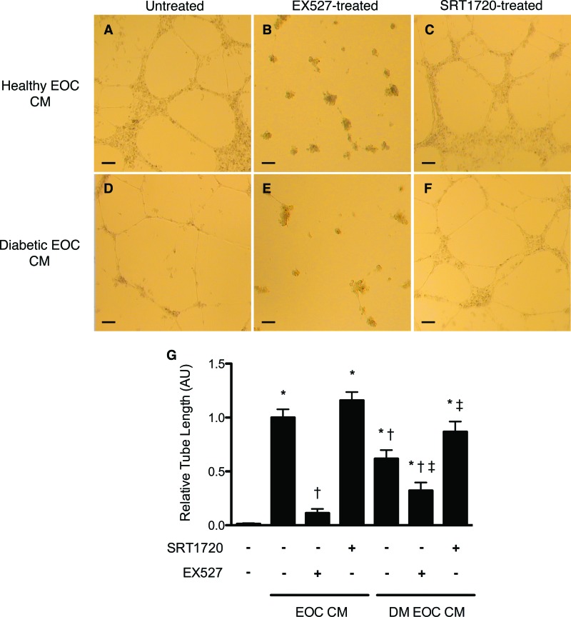
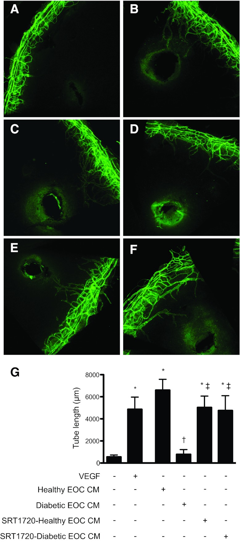
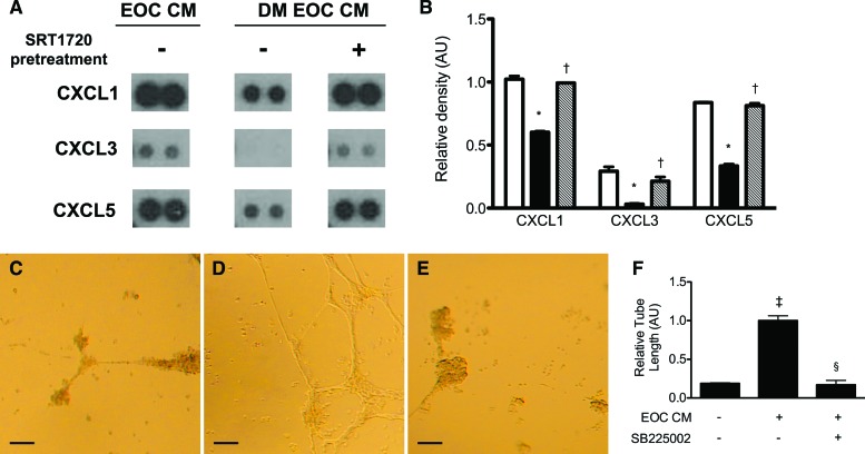
Impaired endothelial repair is a key contributor to microvascular rarefaction and consequent end-organ dysfunction in diabetes. Recent studies suggest an important role for bone marrow-derived early outgrowth cells (EOCs) in mediating endothelial repair, but the function of these cells is impaired in diabetes, as in advanced age. We sought to determine whether diabetes-associated EOC dysfunction might be attenuated by pharmacological activation of silent information regulator protein 1 (SIRT1), a lysine deacetylase implicated in nutrient-dependent life span extension in mammals. Despite being cultured in normal (5.5 mM) glucose for 7 days, EOCs from diabetic rats expressed less SIRT1 mRNA, induced less endothelial tube formation in vitro and neovascularization in vivo, and secreted less of the proangiogenic ELR(+) CXC chemokines CXCL1, CXCL3, and CXCL5. Ex vivo SIRT1 activation restored EOC chemokine secretion and increased the in vitro and in vivo angiogenic activity of EOC conditioned medium derived from diabetic animals to levels similar to that derived from control animals. These findings suggest a pivotal role for SIRT1 in diabetes-induced EOC dysfunction and that its pharmacologic activation may provide a new strategy for the restoration of EOC-mediated repair mechanisms.


Figures

Figure 1.
Figure 1.
Flow cytometric characterization of early outgrowth cells (EOCs). The surface expression of CD34, CD133, and VEGFR2 on EOCs was measured by fluorescence-activated cell sorting. (A–C): Single-marker staining of single-cell EOCs. Left panels: isotype control antibody; right panels: specific antibody. The percentage of positive cells for a given area is listed on each plot. (A): CD34 staining. (B): VEGFR2 staining. (C): CD133 staining. (D): Single-positive CD34+ cells were gated as shown in (A) and examined for VEGFR2 expression. (E): Single-positive CD34+ cells were gated as shown in (A) and examined for CD133 expression. (F): Single-positive CD34+ cells were gated as shown in (A) and examined for staining of VEGFR2 and CD133. Abbreviations: VEGFR2, vascular endothelial growth factor receptor 2; SSC, side scatter.
Figure 2.
Figure 2.
Pharmacologic modulation of silent information regulator protein 1 (SIRT1) activity alters early outgrowth cell (EOC) angiogenic activity in vitro. Although healthy rat EOC CM induced a robust endothelial tube formation response of human umbilical vein endothelial cells seeded in Matrigel (A), diabetic rat EOC CM demonstrated diminished angiogenic activity (D). Inhibition of SIRT1 with EX527 reduced EOC CM angiogenicity, regardless of donor diabetes status (B, E). Activation of SIRT1 with SRT1720 increased the angiogenic activity of diabetic, but not healthy, rat EOC CM (C, F). Quantitative analysis of endothelial tube length is shown (G). *, p < .05 versus serum-free medium; †, p < .05 versus healthy rat EOC CM; ‡, p < .05 versus diabetic rat EOC CM. Images were taken at ×4 magnification. Scale bars = 100 μm. Abbreviations: DM EOC CM, conditioned medium from diabetic rat early outgrowth cells; EOC CM, conditioned medium from healthy rat early outgrowth cells.
Figure 3.
Figure 3.
Silent information regulator protein 1 (SIRT1) activation restores the angiogenic activity of diabetic rat EOC CM in vivo. (A–F): Representative images of corneas harvested from C57BL/6 mice 6 days after injection with 1 μl of phosphate-buffered saline (PBS) (A), 200 ng/μl VEGF (B), CM from healthy rat EOCs (C), CM from diabetic rat EOCs (D), CM from SRT1720-treated healthy rat EOCs (E), or CM from SRT1720-treated diabetic rat EOCs (F). CM was concentrated 40× prior to injection. Six days after injection, the mice were sacrificed, and corneal flat mounts were prepared and stained with an anti-mouse CD31 antibody followed by an Alexa Fluor 488-conjugated secondary antibody (green) to delineate blood vessels. (G): Images were obtained at ×10 magnification, and the cumulative tube lengths of corneal neovessels were quantified. *, p < .05 versus control PBS injection; †, p < .05 versus healthy rat EOC CM injection; ‡, p < .05 versus diabetic rat EOC CM injection. Abbreviations: EOC CM, conditioned medium from healthy rat early outgrowth cells; VEGF, vascular endothelial growth factor.
Figure 4.
Figure 4.
The angiogenic activity of diabetic rat EOC CM is diminished because of a reduction in secretion of proangiogenic ELR+ chemokines. (A): Representative dot blots of EOC CM for the ELR+ chemokines CXCL1, CXCL3, and CXCL5. (B): Quantitative densitometry analysis of the dot blot results. Open bars indicate healthy rat-derived EOC CM. Black bars indicate untreated diabetic rat-derived EOC CM. Cross-hatched bars indicate CM from SRT1720-pretreated diabetic rat-derived EOCs. *, p < .05 versus CM from healthy rat-derived EOCs; †, p < .05 versus untreated diabetic rat-derived EOC CM. (C–F): EOC CM-treated human umbilical vein endothelial cells (HUVECs) were subjected to a Matrigel endothelial tube formation assay, in the presence or absence of SB225002, a selective CXCR2 inhibitor. (C–E): Representative images of endothelial tube formation were taken at ×4 magnification. Scale bars = 100 μm. (C): Serum-free medium-treated HUVECs. (D): Healthy rat-derived EOC CM-treated HUVECs. (E): Healthy rat-derived EOC CM- and SB225002-treated HUVECs. (F): Quantitative analysis of tube length (n = 3). ‡, p < .05 versus serum-free medium; §, p < .05 versus healthy rat-derived EOC CM. Abbreviations: AU, arbitrary units; DM EOC CM, conditioned medium from diabetic rat early outgrowth cells; EOC CM, conditioned medium from healthy rat early outgrowth cells.

References

    1. Loomans CJ, de Koning EJ, Staal FJ, et al. Endothelial progenitor cell dysfunction: A novel concept in the pathogenesis of vascular complications of type 1 diabetes. Diabetes. 2004;53:195–199. - PubMed
    1. Segal MS, Shah R, Afzal A, et al. Nitric oxide cytoskeletal-induced alterations reverse the endothelial progenitor cell migratory defect associated with diabetes. Diabetes. 2006;55:102–109. - PubMed
    1. Fadini GP, Miorin M, Facco M, et al. Circulating endothelial progenitor cells are reduced in peripheral vascular complications of type 2 diabetes mellitus. J Am Coll Cardiol. 2005;45:1449–1457. - PubMed
    1. Potente M, Ghaeni L, Baldessari D, et al. SIRT1 controls endothelial angiogenic functions during vascular growth. Genes Dev. 2007;21:2644–2658. - PMC - PubMed
    1. Yuen DA, Connelly KA, Advani A, et al. Culture-modified bone marrow cells attenuate cardiac and renal injury in a chronic kidney disease rat model via a novel antifibrotic mechanism. PLoS One. 2010;5:e9543. - PMC - PubMed

Publication types

MeSH terms