

Case Reports
doi: 10.1136/bcr-2012-007828.
Unusual presentation of a femoral stress fracture
Affiliations
- PMID: 23283621
- PMCID: PMC3604278
- DOI: 10.1136/bcr-2012-007828
Item in Clipboard
Case Reports
Unusual presentation of a femoral stress fracture
BMJ Case Rep.
.
Abstract
Stress fractures are common injuries in sports medicine. Among these fractures, femoral neck stress fractures frequently have a benign course, especially when it happens in the medial aspect of the neck. This case report describes a stress fracture of the medial aspect of the femoral neck that developed a complete fracture and underwent surgical fixation.
Figures
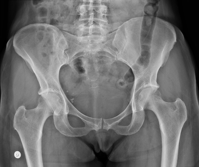
Anteroposterior radiography of the pelvis at initial presentation. This x-ray was considered normal.
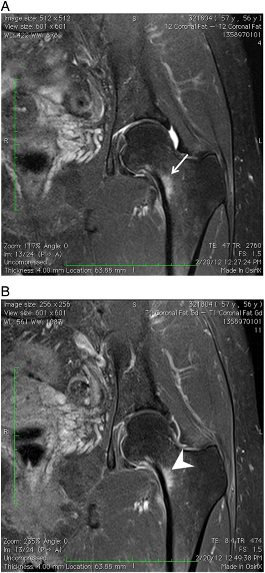
Coronal T2-weighted fat saturated (A) and Coronal T1-weighted fat saturated postgadolinium (B) MR images of left hip demonstrating area of bone marrow oedema adjacent to the medial femoral neck cortex (arrow). Note that the area of bone marrow oedema demonstrates enhancement without focal bone lesion (arrow head). There is also mild soft tissue oedema, without evidence of cortical rupture.
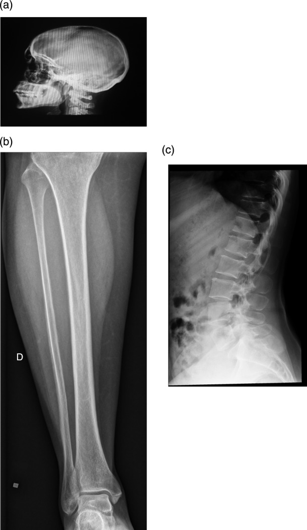
(A) Radiographs obtained for investigation of bone metabolism disorder were considered normal. Lateral skull radiograph. (B) Tibia anteroposterior radiograph. (C) Lumbar spine lateral radiograph.
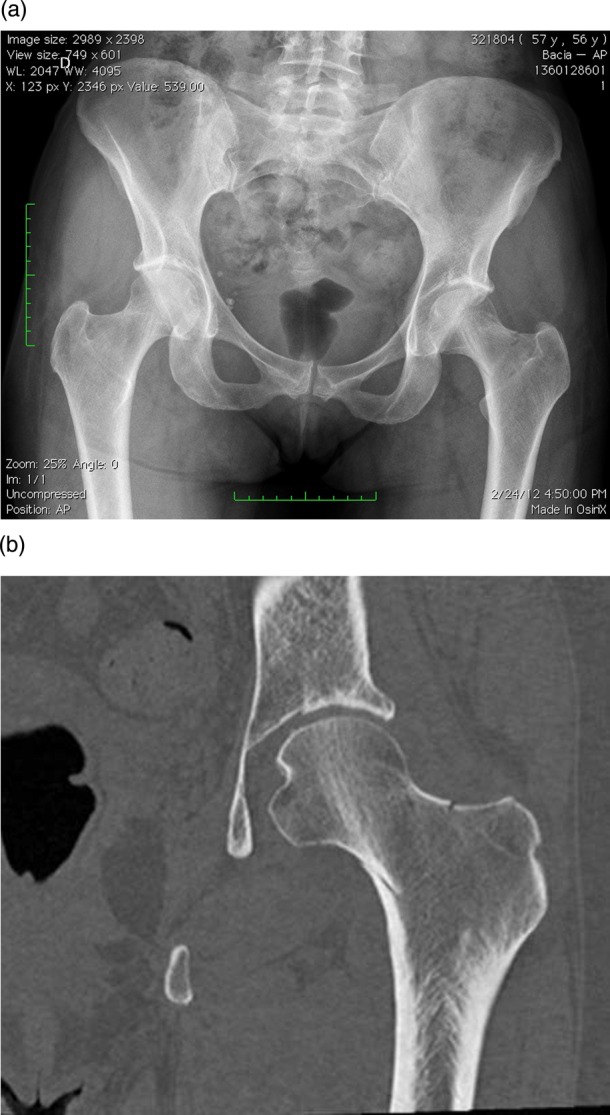
(A) Anteroposterior radiography of the pelvis demonstrating area of cortical thickening of to the left femoral neck cortex with linear lucency suggesting a minimally displaced fracture. (B) Coronal reformat CT image of the left hip demonstrating a minimally displaced oblique fracture of the left femoral neck cortex.
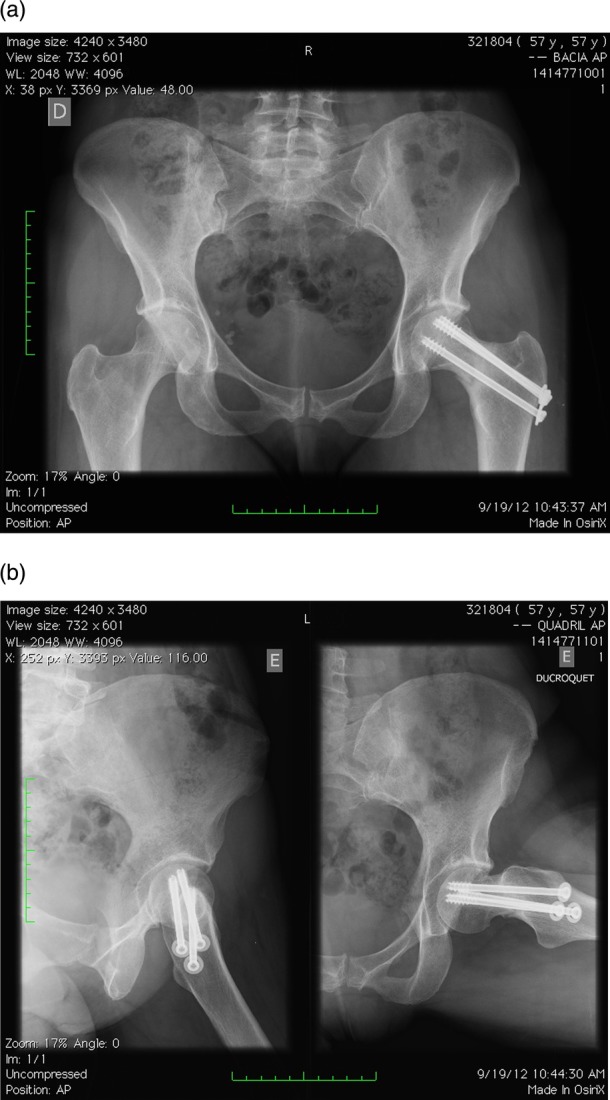
Postoperative radiographs demonstrating three fixation screws within the femoral neck. Note that the femoral neck stress fracture demonstrates callus formation, without deviation.
References
-
- Matheson GO, Clement DB, McKenzie DC, et al. Stress fractures in athletes. A study of 320 cases. Am J Sports Med 1987;15:46–58 - PubMed
-
- Kadel NJ, Teitz CC, Kronmal RA. Stress fractures in ballet dancers. Am J Sports Med 1992;20:445–9 - PubMed
-
- Branch H. March fractures of the femur. J Bone Joint Surg Am 1944;26:387–91
-
- Peterson L. March fracture of the femur—report of a case. J Bone Joint Surg Am 1942;24:185–8
Publication types
MeSH terms
LinkOut - more resources
Full Text Sources
Other Literature Sources
Medical