. 2013 Mar;87(6):3248-60.
doi: 10.1128/JVI.03397-12. Epub 2013 Jan 2.

Insights into head-tailed viruses infecting extremely halophilic archaea

Affiliations

Insights into head-tailed viruses infecting extremely halophilic archaea

Maija K Pietilä et al. J Virol. 2013 Mar.

Abstract

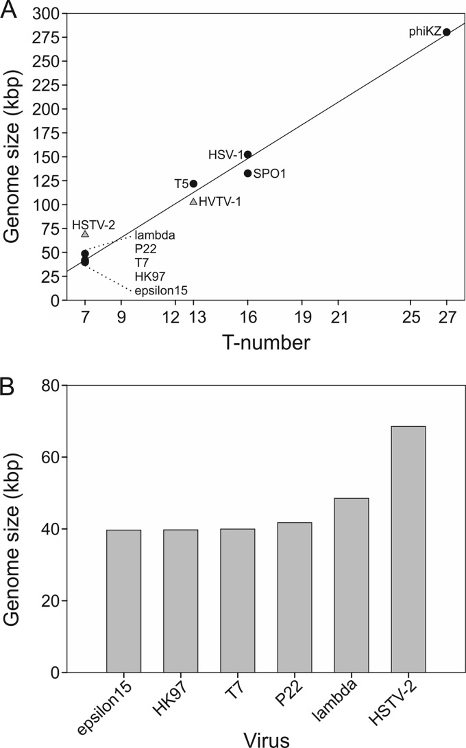
Extremophilic archaea, both hyperthermophiles and halophiles, dominate in habitats where rather harsh conditions are encountered. Like all other organisms, archaeal cells are susceptible to viral infections, and to date, about 100 archaeal viruses have been described. Among them, there are extraordinary virion morphologies as well as the common head-tailed viruses. Although approximately half of the isolated archaeal viruses belong to the latter group, no three-dimensional virion structures of these head-tailed viruses are available. Thus, rigorous comparisons with bacteriophages are not yet warranted. In the present study, we determined the genome sequences of two of such viruses of halophiles and solved their capsid structures by cryo-electron microscopy and three-dimensional image reconstruction. We show that these viruses are inactivated, yet remain intact, at low salinity and that their infectivity is regained when high salinity is restored. This enabled us to determine their three-dimensional capsid structures at low salinity to a ∼10-Å resolution. The genetic and structural data showed that both viruses belong to the same T-number class, but one of them has enlarged its capsid to accommodate a larger genome than typically associated with a T=7 capsid by inserting an additional protein into the capsid lattice.


Figures

Fig 1
Fig 1
Infection cycle of HVTV-1 in its host Haloarcula vallismortis. (A) Turbidity of infected (open circles) and uninfected (closed circles) cultures and number of free viruses in the infected culture (bars). The cultures were washed at 30 min p.i. to remove unadsorbed viruses. Arrows indicate time points of the thin-section samples. (B to D) Micrographs of thin sections of the infected cells observed by transmission electron microscopy at 30 min (B), 10 h (C), and 12 h (D) p.i. Bars, 200 nm (B) and 500 nm (C and D). In panel B, arrows show virions adsorbed to the cell surface. In panels C and D, arrows indicate intracellular genome-containing virions. In panel D, the arrowhead shows an empty capsid.
Fig 2
Fig 2
Viruses are reversibly inactivated under conditions of low salinity without observable morphological changes. (A and B) Effect of lowered ionic strength on the infectivity of HVTV-1 (A) and HSTV-2 (B). The virus stocks (white bars) were diluted either 100-fold (black bars) or 1,000-fold (gray bars) in 23, 18, or 9% salt water (SW) or in 20 mM Tris-HCl (pH 7.2) and incubated for 1 h at 4°C. After incubation, the samples were further diluted in the same buffer, except that the Tris-HCl samples were diluted in 9% SW as well. The diluted samples were incubated for 15 min at RT, and infectivity was then determined. The bars represent the averages of data from two experiments. The infectivity scale in panels A and B is the same, and the infectivities are corrected for the dilution. (C) Cryo-electron micrographs of HVTV-1 at a 4-μm underfocus. (I) Virions incubated in 9% SW; (II) virions incubated in 20 mM Tris-HCl (pH 7.2) buffer; (III) virions which were first incubated in 20 mM Tris-HCl (pH 7.2) and then restored in 9% SW. Bar, 50 nm. (D) Specific infectivity on a logarithmic scale of HVTV-1 virions at different salinities (I to III). (E) Cryo-electron micrographs of HSTV-2 at a 4-μm underfocus showing particles as in panel A. In panel I, the arrow indicates a noncontracted tail, the arrowhead indicates a contracted tail, and the insets show longitudinal views of baseplates dissociated from the capsids. Bar, 50 nm. (F) Specific infectivity on a logarithmic scale of HSTV-2 virions at different salinities (I to III).
Fig 3
Fig 3
Physical map of the HVTV-1 genome. Genes are represented as rectangles, with those above the central scale transcribed from left to right and those below transcribed from right to left, and the markers are spaced at 1-kbp and 100-bp intervals. All predicted genes are protein encoding, with the exception of gene 55, which encodes a tRNAGln. DNA in the virion has a 585-bp direct terminal repeat, indicated by arrows. HNH, histidine-asparagine-histidine homing endonuclease.
Fig 4
Fig 4
Physical map of the HSTV-2 genome. Genes are represented as rectangles above or below the central scale, depending on whether they are transcribed from left to right or right to left, respectively, and the markers are spaced at 1-kbp and 100-bp intervals. All predicted genes are protein encoding, with the exception of gene 70, which encodes a tRNAGln. DNA in the virion has a 340-bp direct terminal repeat, indicated by arrows.
Fig 5
Fig 5
HVTV-1 and HSTV-2 structural proteins. (A) Protein pattern of virions in a Tricine-SDS-polyacrylamide gel stained with Coomassie blue. 1× indicates virions purified in a sucrose gradient by rate-zonal centrifugation, and 2× indicates virions further purified in a CsCl gradient by equilibrium centrifugation. Numbers on the left indicate molecular mass markers. (B) Amino acid sequences of the major structural proteins. The N-terminal sequence determined from the protein band is shown by boldface type and underlining, except that the sixth amino acid in HSTV-2 gp20 could not be identified. The theoretical molecular masses of the mature proteins are given in parentheses.
Fig 6
Fig 6
Cryo-EM reconstruction of the HVTV-1 and HSTV-2 capsids. (A and B) Central section of HVTV-1 (A) and HSTV-2 (B) capsids. The icosahedral reconstructions are viewed along a 2-fold axis. The ellipse, triangle, and pentagon indicate 2-fold-, 3-fold-, and 5-fold-symmetry axes, respectively. Bar, 10 nm. (C and D) Isosurface representation of HVTV-1 (C) and HSTV-2 (D) capsid reconstructions along an icosahedral 2-fold axis. The capsids are displayed at a threshold of a sigma value of 3.0. The symmetry axes are shown as in panel A. Coloring was done using radial-depth cueing (bar, 325- to 425-Å radius) in UCSF Chimera (54).
Fig 7
Fig 7
Genome size and T-number comparison. (A) Genome size versus T-number. Triangles indicate HSTV-2 (GenBank accession number KC117376) and HVTV-1 (accession number KC117377), accordingly. Circles indicate epsilon15 (accession number AY150271), HK97 (accession number AF069529), T7 (accession number V01146), P22 (accession number BK000583), lambda (accession number J02459), T5 (accession number AY543070), SPO1 (accession number FJ230960), HSV-1 (accession number X14112), and phiKZ (accession number AF399011). (B) Genome sizes of T=7 head-tailed viruses.
Fig 8
Fig 8
Organization of the two capsid proteins of HSTV-2, illustrated for two icosahedral facets. Shown is the capsid protein organization of bacteriophage lambda (structure under EMDB accession number EMD-5012 filtered to a 9.8-Å resolution and isosurface drawn at a threshold of a sigma value of 3.0) (73) (top) and archaeal virus HSTV-2 (isosurface drawn at a threshold of a sigma value of 3.0) (bottom). For two icosahedral facets, positions of MCP pentamers and hexamers are shown in blue, and positions of minor capsid protein trimers are shown in red. The magnified region shows cryo-EM density of the HSTV-2 minor capsid protein trimer with the bacteriophage lambda gpD protein structure (PDB accession number 1C5E) (74) fitted in and shown as a red ribbon. Visualization was done by using UCSF Chimera (54). Fitting of the gpD structure was done with the “fit in map” tool in UCSF Chimera.

References

    1. Chaban B, Ng SYM, Jarrell KF. 2006. Archaeal habitats—from the extreme to the ordinary. Can. J. Microbiol. 52:73–116 - PubMed
    1. DeLong EF, Pace NR. 2001. Environmental diversity of bacteria and archaea. Syst. Biol. 50:470–478 - PubMed
    1. Pina M, Bize A, Forterre P, Prangishvili D. 2011. The archeoviruses. FEMS Microbiol. Rev. 35:1035–1054 - PubMed
    1. Ackermann HW, Prangishvili D. 2012. Prokaryote viruses studied by electron microscopy. Arch. Virol. 157:1843–1849 - PubMed
    1. Atanasova NS, Roine E, Oren A, Bamford DH, Oksanen HM. 2012. Global network of specific virus-host interactions in hypersaline environments. Environ. Microbiol. 14:426–440 - PubMed

Publication types

MeSH terms

Associated data

LinkOut - more resources