. 2013 Mar;87(6):3108-18.
doi: 10.1128/JVI.02826-12. Epub 2013 Jan 2.

Identification of the N-terminal domain of the influenza virus PA responsible for the suppression of host protein synthesis

Affiliations

Identification of the N-terminal domain of the influenza virus PA responsible for the suppression of host protein synthesis

Emily A Desmet et al. J Virol. 2013 Mar.

Abstract

Cellular protein synthesis is suppressed during influenza virus infection, allowing for preferential production of viral proteins. To explore the impact of polymerase subunits on protein synthesis, we coexpressed enhanced green fluorescent protein (eGFP) or luciferase together with each polymerase component or NS1 of A/California/04/2009 (Cal) and found that PA has a significant impact on the expression of eGFP and luciferase. Comparison of the suppressive activity on coexpressed proteins between various strains revealed that avian virus or avian-origin PAs have much stronger activity than human-origin PAs, such as the one from A/WSN/33 (WSN). Protein synthesis data suggested that reduced expression of coexpressed proteins is not due to PA's reported proteolytic activity. A recombinant WSN containing Cal PA showed enhanced host protein synthesis shutoff and induction of apoptosis. Further characterization of the PA fragment indicated that the N-terminal domain (PANt), which includes the endonuclease active site, is sufficient to suppress cotransfected gene expression. By characterizing various chimeric PANts, we found that multiple regions of PA, mainly the helix α4 and the flexible loop of amino acids 51 to 74, affect the activity. The suppressive effect of PANt cDNA was mainly due to PA-X, which was expressed by ribosomal frameshifting. In both Cal and WSN viruses, PA-X showed a stronger effect than the corresponding PANt, suggesting that the unique C-terminal sequences of PA-X also play a role in suppressing cotransfected gene expression. Our data indicate strain variations in PA gene products, which play a major role in suppression of host protein synthesis.


Figures
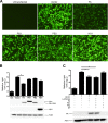
Fig 1
Fig 1
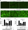
PA reduces the expression of cotransfected proteins. 293T cells were cotransfected with expression vectors containing eGFP or luciferase genes together with the indicated Cal genes. (A) eGFP expression at 24 h posttransfection. (B) Luciferase expression from pRL-SV40 in cells cotransfected with the indicated Cal genes in pCAGGS. Expressed viral proteins were determined by Western blotting using an anti-Flag Ab. Anti-β-actin Ab was used for a loading control. (C) Luciferase expression in cells cotransfected with the indicated Cal polymerase subunits. Cells were transfected with 0.4 μg of each subunit (supplemented with empty vector) to total 1.6 μg. PA expression was determined by Western blotting using anti-PA antibody. *, P < 0.05.
Fig 2
Fig 2
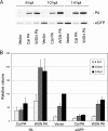
Avian-origin PAs are more active in suppressing coexpressed proteins. 293T cells were cotransfected with expression vectors containing eGFP or luciferase genes together with the PA genes of the indicated viruses. (A) eGFP expression as measured at 24 h posttransfection. (B) Luciferase expression in 293T cells as measured in cells transfected with the indicated genes in pCAGGS together with pRL-SV40. Expressed PA was determined by Western blotting using anti-Flag Ab. (C) Suppression of luciferase expression by various PAs in DF-1 cells. *, P < 0.05.
Fig 3
Fig 3
Cal PA strongly suppresses production of coexpressed nonviral proteins. (A) 293T cells were cotransfected with expression vectors containing eGFP and either WSN PA, Cal PA, or empty vector. At the indicated time points following transfection (hpt), cells were labeled with [35S]Met-Cys for 30 min, and eGFP and PA were immunoprecipitated using an anti-Flag Ab. (B) The relative volumes of eGFP and PA proteins at each time point were quantified using Quantity One 1-D analysis software (Bio-Rad).
Fig 4
Fig 4
Virus growth and suppression of cellular protein synthesis. (A) Reduced growth of WSN-CalPA in A549 cells. A549 cells were infected with either WSN or WSN-CalPA at an MOI of 0.01 and cultured in the presence of trypsin at 1 μg/ml for 96 h. Virus titers in the culture supernatants were measured. (B and C) WSN-CalPA inhibits cellular protein synthesis more efficiently than WSN. A549 cells were either left uninfected or infected with WSN or WSN-CalPA at an MOI of 3. At the indicated times after infection, cells were labeled with [35S]Met-Cys for 30 min and total cell lysates were resolved by SDS-PAGE (B). Representative cellular proteins above 100 kDa (CP1) and a 50-kDa band (CP2) as well as viral proteins (NP and NS1) were quantified using Quantity One 1-D Analysis Software (C).
Fig 5
Fig 5
Enhanced apoptosis induction by WSN-CalPA. A549 cells were infected with either WSN or WSN-CalPA at an MOI of 3, and cell viability and apoptotic state were determined. (A) Cell viability was determined by trypan blue staining. (B) Apoptotic cells were identified at 12 and 24 h postinfection. Annexin V (green) and propidium iodide (PI) (red) signals were visualized using a fluorescence microscope.
Fig 6
Fig 6
The N-terminal domain of PA determines shutoff activity. (A) Schematic diagram of the chimeric PA constructs. (B) Luciferase expression from pRL-SV40 in cells cotransfected with the indicated chimeric PA genes in pCAGGS. (C) Expression of PA proteins was determined by Western blotting using an anti-Flag Ab. Anti-β-actin Ab was used for a loading control.
Fig 7
Fig 7
The N-terminal 257 residues are sufficient to induce shutoff. (A) Luciferase expression from pRL-SV40 in cells cotransfected with the indicated PA genes in pCAGGS. PANt indicates a protein containing the PA N-terminal 257 residues, and PACt indicates a protein containing the PA C-terminal residues 258 to 716. (B) Expression of the PA fragments was determined by Western blotting using an anti-Flag Ab. Anti-β-actin Ab was used as a loading control.
Fig 8
Fig 8
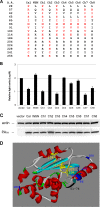
Identification of the residues required for shutoff. (A) Sequence differences between the Cal and WSN PA N-terminal regions (residues 1 to 257). The amino acid differences in the Cal/WSN chimeric PA N-terminal constructs are also shown. (B) Luciferase expression from pCAGGS-Luc in cells cotransfected with the indicated chimeric PANt genes in pCAGGS. (C) Expression of chimeric PANt was determined by Western blotting using an anti-Flag Ab. Anti-β-actin Ab was used as loading control. (D) Crystal structure of the PA N-terminal domain, highlighting the locations of residues that affect shutoff activity (blue). Residues in the flexible domain with an undetermined structure are shown in black. The putative endonuclease active site (P107D108X10E119K134) (29) is shown in yellow.
Fig 9
Fig 9
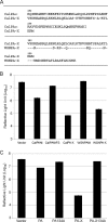
Shutoff activity of PA-X. (A) Sequence differences in the unique region between the Cal PA N-terminal fragment and PA-X (top). The unique C-terminal region of PA-X is highly homologous between Cal and WSN, except that WSN contains an additional 20 residues (bottom). (B) Luciferase expression from pCAGGS-Luc in cells cotransfected with the indicated PANt and PA-X genes in pCAGGS. Cells were cultured for 20 h after transfection. (C) Shutoff activities of CalPA134A and CalPA-X134A were determined as described for panel B except that transfected cells were cultured for 24 h.

References

    1. Smith NM, Bresee JS, Shay DK, Uyeki TM, Cox NJ, Strikas RA. 2006. Prevention and control of influenza: recommendations of the Advisory Committee on Immunization Practices (ACIP). MMWR Recommend. Rep. 55:1–42 - PubMed
    1. Perrone LA, Plowden JK, Garcia-Sastre A, Katz JM, Tumpey TM. 2008. H5N1 and 1918 pandemic influenza virus infection results in early and excessive infiltration of macrophages and neutrophils in the lungs of mice. PLoS Pathog. 4:e1000115 doi:10.1371/journal.ppat.1000115 - DOI - PMC - PubMed
    1. Katze MG, DeCorato D, Krug RM. 1986. Cellular mRNA translation is blocked at both initiation and elongation after infection by influenza virus or adenovirus. J. Virol. 60:1027–1039 - PMC - PubMed
    1. Katze MG, Detjen BM, Safer B, Krug RM. 1986. Translational control by influenza virus: suppression of the kinase that phosphorylates the alpha subunit of initiation factor eIF-2 and selective translation of influenza viral mRNAs. Mol. Cell. Biol. 6:1741–1750 - PMC - PubMed
    1. Garcia-Sastre A, Egorov A, Matassov D, Brandt S, Levy DE, Durbin JE, Palese P, Muster T. 1998. Influenza A virus lacking the NS1 gene replicates in interferon-deficient systems. Virology 252:324–330 - PubMed

Publication types

MeSH terms