. 2013 Feb;24(4):510-20.
doi: 10.1091/mbc.E12-06-0492. Epub 2013 Jan 2.

Visualization of the exocyst complex dynamics at the plasma membrane of Arabidopsis thaliana

Affiliations

Visualization of the exocyst complex dynamics at the plasma membrane of Arabidopsis thaliana

Matyás Fendrych et al. Mol Biol Cell. 2013 Feb.

Abstract

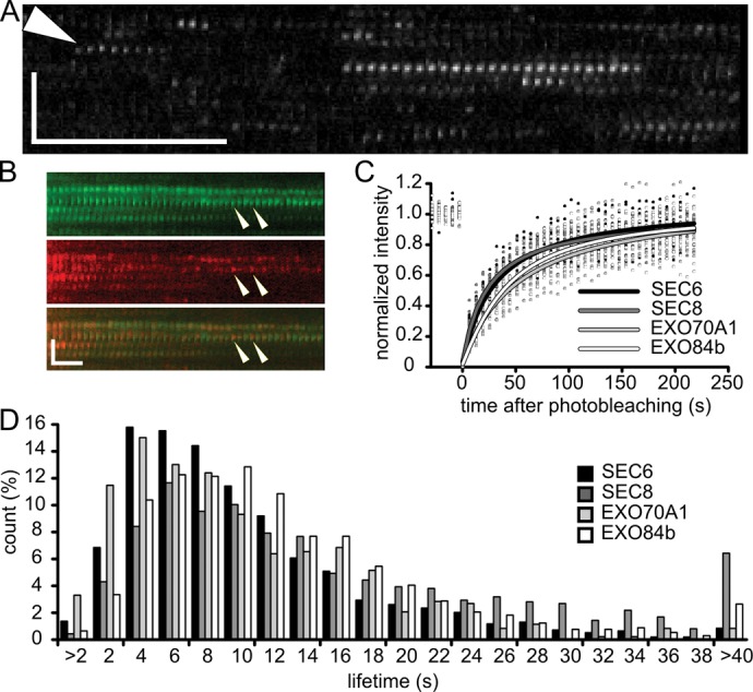
The exocyst complex, an effector of Rho and Rab GTPases, is believed to function as an exocytotic vesicle tether at the plasma membrane before soluble N-ethylmaleimide-sensitive factor attachment protein receptor (SNARE) complex formation. Exocyst subunits localize to secretory-active regions of the plasma membrane, exemplified by the outer domain of Arabidopsis root epidermal cells. Using variable-angle epifluorescence microscopy, we visualized the dynamics of exocyst subunits at this domain. The subunits colocalized in defined foci at the plasma membrane, distinct from endocytic sites. Exocyst foci were independent of cytoskeleton, although prolonged actin disruption led to changes in exocyst localization. Exocyst foci partially overlapped with vesicles visualized by VAMP721 v-SNARE, but the majority of the foci represent sites without vesicles, as indicated by electron microscopy and drug treatments, supporting the concept of the exocyst functioning as a dynamic particle. We observed a decrease of SEC6-green fluorescent protein foci in an exo70A1 exocyst mutant. Finally, we documented decreased VAMP721 trafficking to the plasma membrane in exo70A1 and exo84b mutants. Our data support the concept that the exocyst-complex subunits dynamically dock and undock at the plasma membrane to create sites primed for vesicle tethering.


Figures

FIGURE 1:
FIGURE 1:
Exocyst subunits colocalize in distinct foci at the plasma membrane. (A) CLSM section of Arabidopsis root expressing EXO84b-GFP. The signal decorates outer epidermal PM. In recently divided cells, the exocyst is focused on the maturing cell walls (asterisk). (B) Exocyst subunits form distinct foci at the PM when observed by VAEM. Foci appearance is shown in the PM of outer epidermal root cells in elongation (left) and root hair zones (right). Scale bars, 10 μm. (C) Exocyst foci density in root epidermal cells; n ≥ 10 cells for each column; error bars, SDs. (D) Quantification (%) of the exocyst subunits and DRP1C colocalizations in Arabidopsis root epidermal cells. Pairs indicated in E were evaluated. Green, red, and yellow colors represent GFP only, mRFP only, and their colocalization, respectively. (E) Localization of SEC6, SEC8, and EXO84b exocyst subunits. Green, red, and yellow circles denote GFP-only, mRFP-only, and GFP- and mRFP-positive foci, respectively. Scale bars, 1 μm.
FIGURE 2:
FIGURE 2:
Exocyst foci dynamics. (A) A kymographic representation of EXO84b-GFP at the PM of an elongating root epidermal cell. Arrowhead points to lateral movement of EXO84b-GFP in the vertical direction. Horizontal and vertical bars represent 5 s and 3 μm, respectively. (B) A kymographic representation of SEC6-GFP and EXO84b-mRFP colocalization in the exocyst foci. Recruitment of new EXO84b-mRFP molecules is apparent (arrowheads). The horizontal and vertical bars represent 10 s and 2 μm, respectively. (C) FRAP of exocyst subunits at lateral PM of elongating root epidermal cells. Each curve was constructed from ≥10 FRAP experiments; the individual measurements are represented by dots. (D) Histogram showing distribution of exocyst foci lifetimes measured in kymographs; n ≥ 600 foci for each subunit.
FIGURE 3:
FIGURE 3:
EXO84b-GFP and cytoskeleton in root epidermal cells. (A) EXO84B-GFP (green) does not colocalize with actin as visualized by Lifeact-mRFP (red). Short-term actin disruption (10 min, right) does not influence the appearance of exocyst foci. Scale bars, 2 μm. (B) Localization of EXO84b-mRFP (red) and microtubules (green, MAP4-GFP). After 10-min APM treatment, microtubules are disrupted, whereas exocyst foci remain unaffected. Scale bars, 2 μm. (C, D) EXO84b-GFP foci 1 h after disruption of actin (C) and microtubule (D) cytoskeleton. Scale bars, 5 μm. (E, F) EXO84b-GFP in CLSM root sections 1 h after disruption of actin (E) and control (F). Hyperpolarization of the exocyst signal is obvious in E. Scale bars, 10 μm. (G, H) FRAP curves demonstrate retarded recovery of EXO84b-GFP at the PM of the epidermal root cells upon 1-h treatment by latB (G). MT disruption had no effect. Error bars, SDs; n = 12 (controls), 20 (latB and APM treatments).
FIGURE 4:
FIGURE 4:
Colocalization of exocyst foci with vesicle marker and electron microscopy analysis of the lateral plasma membrane of root epidermal cells. (A–C) Comparison of VAMP721 (A), exocyst foci (B), and SYP132-GFP (C) CLSM localization (left) and dynamics visualized by VAEM (right). GFP-VAMP721 localizes to epidermal PM and endosomes and EXO84b-GFP signal is prominent at the outer epidermal PM, whereas SYP132-GFP is evenly distributed along the entire PM; the seemingly stronger signal of the intercellular membranes results from summing of the adjacent PM signal. Kymographs demonstrate that VAMP721 and exocyst label PM-localized foci with similar appearance (A, B). VAMP721 foci dwelling at the PM are preceded by movement of the foci (arrowheads). Motile foci and endosomes are visible in the kymograph (B). SYP132-GFP (C) is highly dynamic compared with EXO84b-labeled foci. Scale bars, 5 μm (left); horizontal and vertical bars (right) represent 5 s and 3 μm, respectively. (D) Colocalization of GFP-VAMP721 and EXO84b-mRFP foci (scale bar, 2 μm), and quantification (%) of the colocalization (left). The random overlap quantification is shown on the right. Note the weak PM signal of GFP-VAMP721 apart from the larger foci. (E) Kymographic representation of the colocalization between GFP-VAMP721 and EXO84b-mRFP; horizontal and vertical bars represent 10 s and 2 μm, respectively. (F, G) HPF-AFS electron microscopy analysis of the lateral PM of Arabidopsis root epidermal cells. (H) Table summarizing the length and area of lateral PM analyzed and number of visible vesicles. From the actual vesicle number observed it is clear that only a subset of exocyst foci are tethering a vesicle. Numerous Golgi and endomembranes are apparent in F, but no vesicles are tethered below the lateral PM. In the lower cell, paramural bodies are present (arrowhead). An example of vesicle tethered at the PM is shown in G; note also the presence of numerous vesicles in the cytoplasm close to the Golgi. (I) Magnified inset from (G); the distance of the vesicle from the PM is 23 nm. Scale bars, 1 μm in F and G and 100 nm in I.
FIGURE 5:
FIGURE 5:
The exocyst is insensitive to brefeldin A treatment. Plants expressing exocyst subunits and VAMP721 GFP fusions were treated with BFA for 2 h as indicated. The exocyst subunits remained localized on the lateral PM in the root epidermal cells, whereas the VAMP721 aggregated in BFA compartments. Scale bars, 10 μm.
FIGURE 6:
FIGURE 6:
Mutations in exocyst subunits decrease the incidence of exocyst foci at the PM and lead to a decrease in exocytosis. (A) SEC6-GFP exocyst foci in wild-type and exo70A1-mutant root epidermal cells; scale bars, 2 μm. (B) quantification of SEC6-GFP foci density in exo70A1 wild-type and mutant cells; error bars, SDs; n = 23 and 12 cells for exo70A1mm and exo70A1 wild type, respectively. (C) GFP-VAMP721 localizes to the lateral PM and endomembranes in the wild type and exo70A1 phenotypic wild-type cells. In exo70A1 and exo84b1 exocyst mutants, GFP-VAMP721 localization to the lateral PM decreases, and, in turn, the signal of endomembrane compartment increases. Calibration bar of the color coding is shown on the right; scale bars, 5 μm. (D) Quantification of the lateral PM domain/cytoplasmic signal intensity ratio in wild-type, exo70A1 wild-type, and exo70A1 mutants. The exo84b mutants were not quantified due to the lack of signal at the PM. n = 48 cells in 18 seedlings (wild type), n = 54 cells in 18 seedlings (exo70A1 wild type), and n = 45 cells in 16 seedlings (exo70A1 mutants); error bars, SDs.

Similar articles

Cited by

References

    1. Allersma MW, Wang L, Axelrod D, Holz RW. Visualization of regulated exocytosis with a granule-membrane probe using total internal reflection microscopy. Mol Biol Cell. 2004;15:4658–4668. - PMC - PubMed
    1. Barg S, Knowles M, Chen X, Midorikawa M, Almers W. Syntaxin clusters assemble reversibly at sites of secretory granules in live cells. Proc Natl Acad Sci USA. 2010;107:20804–20809. - PMC - PubMed
    1. Batoko H, Zheng HQ, Hawes C, Moore I. A rab1 GTPase is required for transport between the endoplasmic reticulum and Golgi apparatus and for normal Golgi movement in plants. Plant Cell. 2000;12:2201–2218. - PMC - PubMed
    1. Battey N, James N, Greenland A, Brownlee C. Exocytosis and endocytosis. Plant Cell. 1999;11:643–660. - PMC - PubMed
    1. Bendezú FO, Martin SG. Actin cables and the exocyst form two independent morphogenesis pathways in the fission yeast. Mol Biol Cell. 2010;22:44–53. - PMC - PubMed

Publication types

MeSH terms