. 2013 Mar;24(5):643-58.
doi: 10.1091/mbc.E12-09-0659. Epub 2013 Jan 2.

Rab11-family interacting proteins define spatially and temporally distinct regions within the dynamic Rab11a-dependent recycling system

Affiliations

Rab11-family interacting proteins define spatially and temporally distinct regions within the dynamic Rab11a-dependent recycling system

Nicholas W Baetz et al. Mol Biol Cell. 2013 Mar.

Abstract

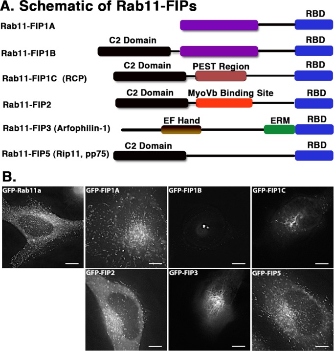
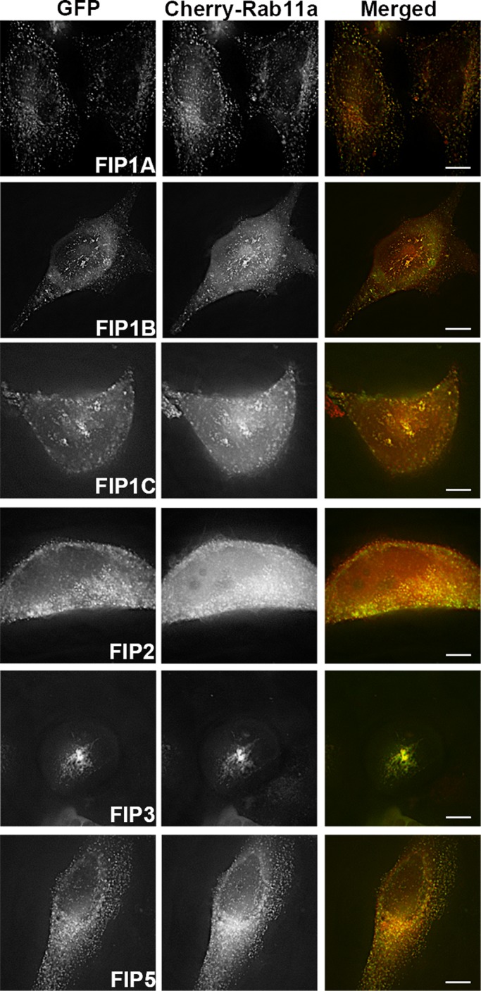
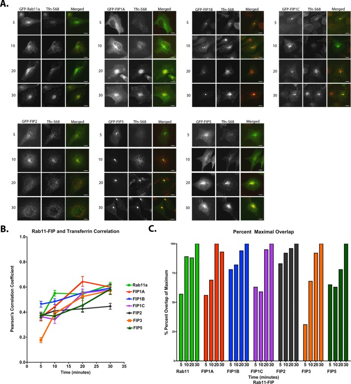
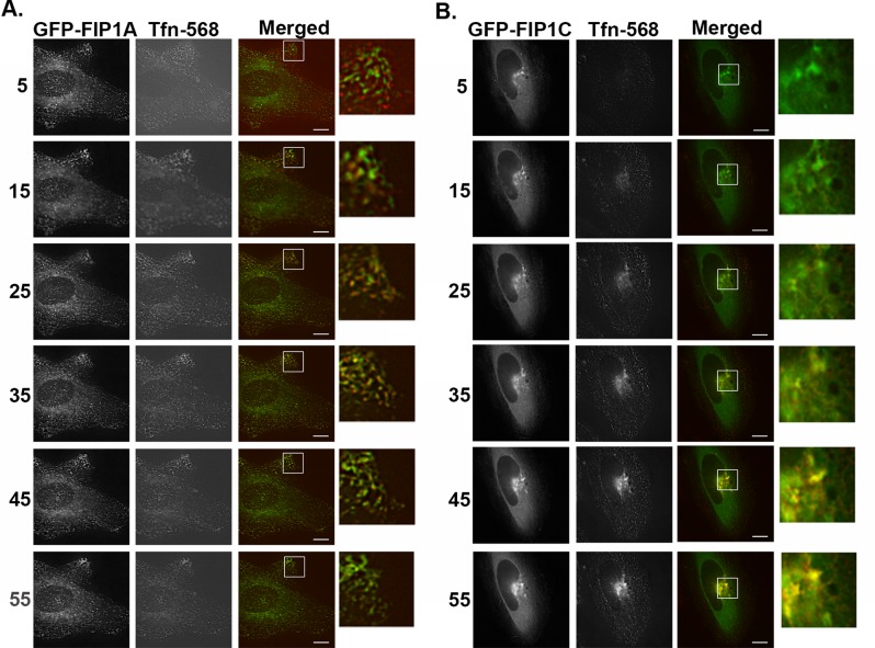
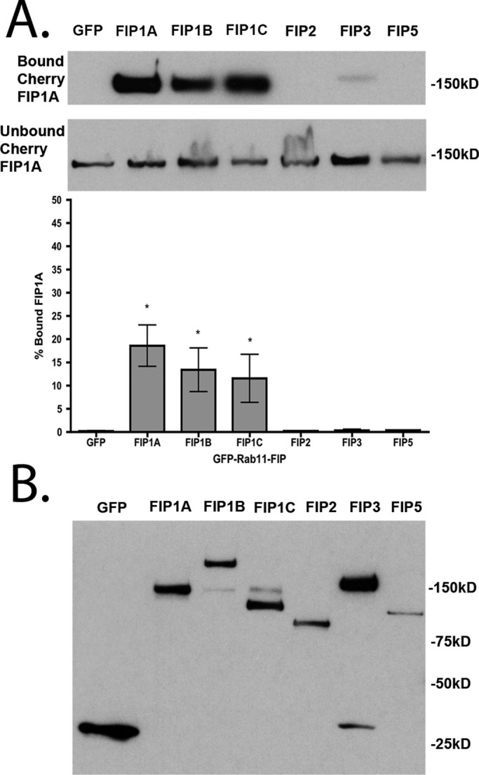
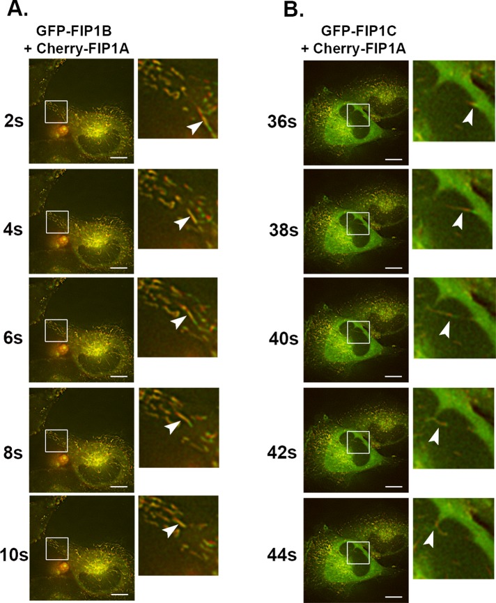
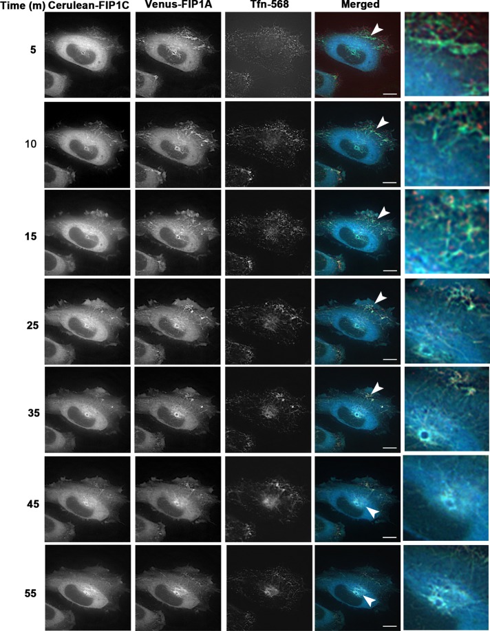
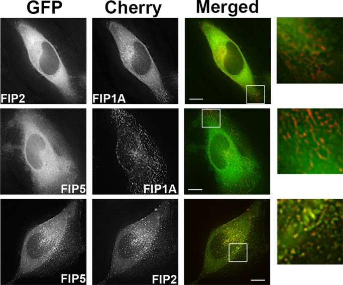
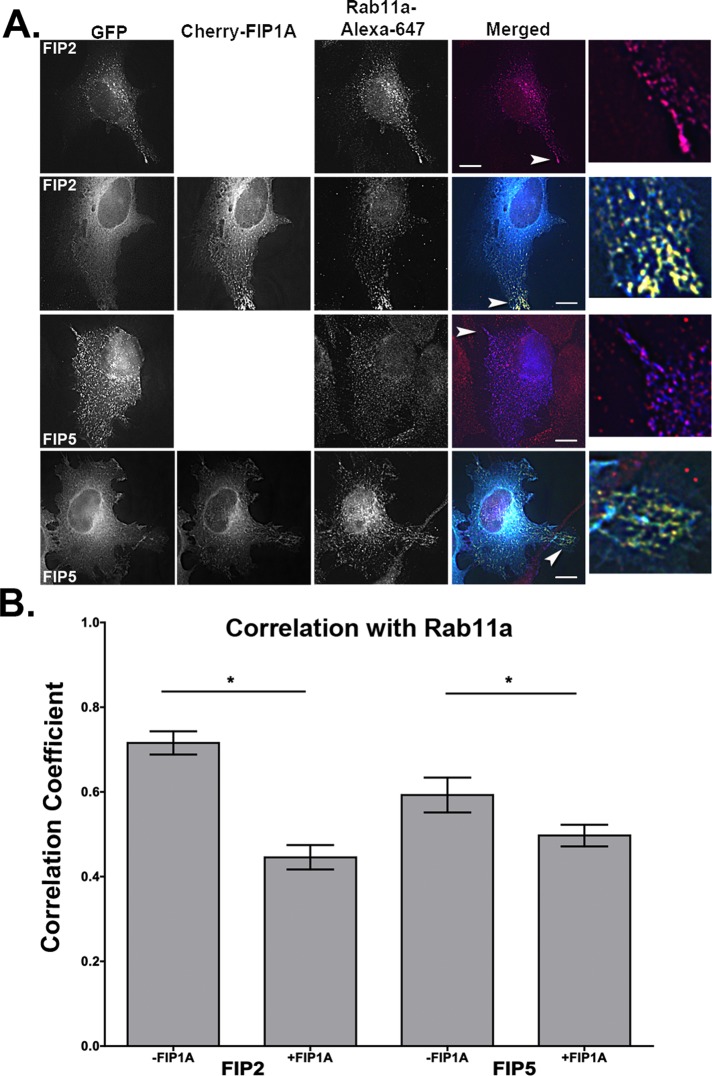
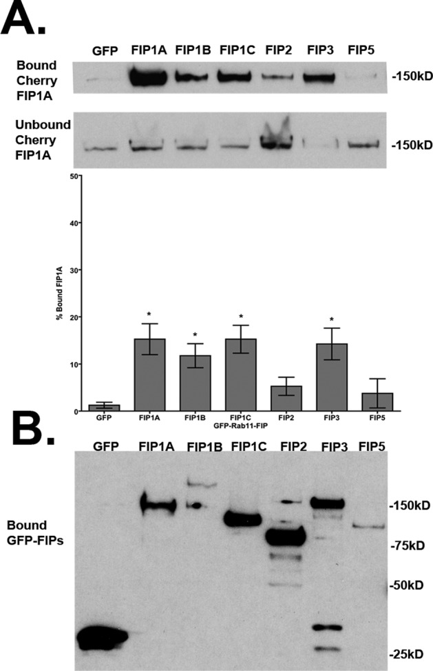
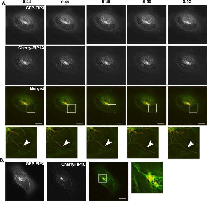
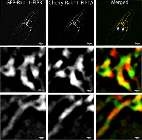
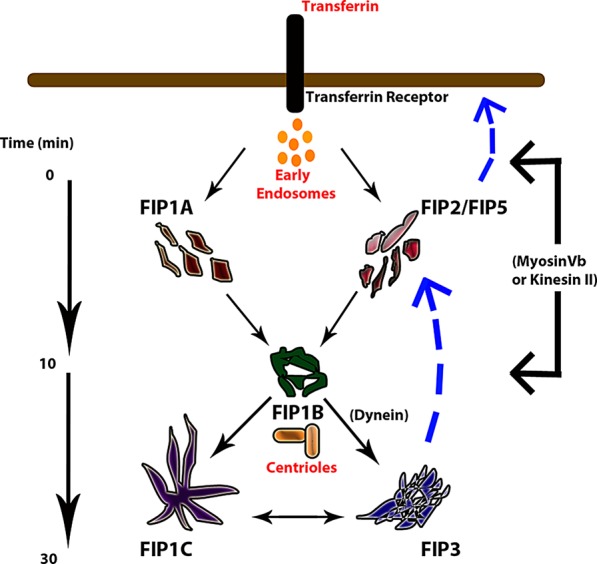
The Rab11-family interacting proteins (Rab11-FIPs) facilitate Rab11-dependent vesicle recycling. We hypothesized that Rab11-FIPs define discrete subdomains and carry out temporally distinct roles within the recycling system. We used live-cell deconvolution microscopy of HeLa cells expressing chimeric fluorescent Rab11-FIPs to examine Rab11-FIP localization, transferrin passage through Rab11-FIP-containing compartments, and overlap among Rab11-FIPs within the recycling system. FIP1A, FIP2, and FIP5 occupy widely distributed mobile tubules and vesicles, whereas FIP1B, FIP1C, and FIP3 localize to perinuclear tubules. Internalized transferrin entered Rab11-FIP-containing compartments within 5 min, reaching maximum colocalization with FIP1B and FIP2 early in the time course, whereas localization with FIP1A, FIP1C, FIP3, and FIP5 was delayed until 10 min or later. Whereas direct interactions with FIP1A were only observed for FIP1B and FIP1C, FIP1A also associated with membranes containing FIP3. Live-cell dual-expression studies of Rab11-FIPs revealed the tubular dynamics of Rab11-FIP-containing compartments and demonstrated a series of selective associations among Rab11-FIPs in real time. These findings suggest that Rab11-FIP1 proteins participate in spatially and temporally distinct steps of the recycling process along a complex and dynamic tubular network in which Rab11-FIPs occupy discrete domains.


Figures

FIGURE 1:
FIGURE 1:
EGFP–Rab11-FIPs in live HeLa cells occupy distinct compartments. (A) Schematic representations of the Rab11-FIPs examined in the present study, the names used for each of the proteins in this and other studies, and a general outline of previously characterized regions within these proteins. Time-lapse images of each Rab11-FIP condition were collected, and representative single frames are presented for each condition in B. Corresponding time-lapse videos for the Rab11-FIP1 proteins and Rab11a are presented in Supplemental Video S1. Two general phenotypic differences were observed in FIP localization and movement. FIP1A, FIP2, and FIP5 maintain a wide distribution, similar to that observed for Rab11a. FIP1B, FIP1C, and FIP3 are primarily more centralized in localization. In addition, FIP1C and FIP3 displayed evidence of branching tubules emanating from the central perinuclear region. Data represent at least three independent experiments. Bars, 10 μm.
FIGURE 2:
FIGURE 2:
Rab11a in live HeLa cells is targeted differently in the presence of Rab11-FIPs. Single frames were collected from time-lapse imaging of Rab11-FIPs with Rab11a. FIP1A (Supplemental Video S2), FIP2, and FIP5 overlapped with Rab11a throughout the cell, whereas FIP1B and FIP1C (Supplemental Video S3), as well as FIP3, accumulate Rab11a in more centralized compartments. Data represent at least three independent experiments. Bars, 10 μm.
FIGURE 3:
FIGURE 3:
EGFP–Rab11-FIPs in HeLa cells overlap with transferrin at temporally distinct points. HeLa cells transfected with EGFP–Rab11-FIPs were incubated with transferrin–Alexa 568 for 5, 10, 20, and 30 min and imaged to examine overlap. FIP1B demonstrated the most overlap at 5 min and reached ∼80% of overlap during this time. FIP2 also exhibited 80% of overlap in 5 min as well, whereas FIP1A, FIP1C, FIP3, and FIP5 displayed less overlap with transferrin at 5 min, and yet the progression of overlap toward the maximum values within this group was staggered between10 and 20 min. FIP1A progressed to maximum overlap at 20 min, whereas FIP1C, FIP3, and FIP5 showed a delayed rise to maximum overlap at 30 min. (A) Representative images for each time point (25 cells) in each condition (100 cells). (B, C) Average Pearson's r (also see Tables 1 and 2) at each time point and percentage maximum overlap calculated from the average values. Results are presented as mean ± SEM. Bars,10 μm.
FIGURE 4:
FIGURE 4:
Transferrin traffics through FIP1A and FIP1C at temporally distinct time points. Live HeLa cells transfected with either EGFP-FIP1A or EGFP-FIP1C were incubated with transferrin–Alexa 568 for 5 min and then chased for 1 h. Evidence of overlap between transferrin and FIP1A (Supplemental Video S4) was observed as early as 5 min, and transferrin signal returns to baseline by 45 min (A). FIP1C (Supplemental Video S4) overlap with transferrin is evident at 15 min and persists until 55 min (B). Imaging of transferrin overlap with EGFP-FIPs was carried out on a DeltaVision deconvolution microscope. Time points (minutes) are indicated to the left of the images. Data represent at least three independent experiments. Bars,10 μm.
FIGURE 5:
FIGURE 5:
FIP1A is isolated specifically with FIP1A, FIP1B, and FIP1C in the presence of 1% Triton X-100. (A) Western blots of the bound and unbound FIP1A isolated with EGFP-FIPs from 1% Triton X-100–solubilized HEK293 cell preparations. Significant percentages of FIP1A were recovered with FIP1A (18.59 ± 4.45), FIP1B (13.40 ± 4.69), and FIP1C (11.55 ± 5.15) in comparison to the EGFP control (0.20 ± 0.10; p = 0.05). Bound material is presented as a percentage of total material recovered. (B) EGFP-FIPs isolated in each condition. Data represent at least three independent experiments. Results are presented as mean ± SEM.
FIGURE 6:
FIGURE 6:
FIP1A overlaps with FIP1B and FIP1C along tubular compartments. Time-lapse imaging of mCherry-FIP1A with EGFP-FIP1B (Supplemental Video S5) or EGFP-FIP1C (Supplemental Video S6) in live HeLa cells conducted over 2 min. (A) FIP1B on tubular compartments with FIP1A. The insets show an example tubule compartment labeled with both mCherry-FIP1A and EGFP-FIP1B moving from the perinuclear region to the periphery of the cell. (B) EGFP-FIP1C and mCherry-FIP1A along tubular compartments that move in the same direction and change directions together. Insets highlight the overlap along the compartment and the movement of an example tubule. Data represent at least three independent experiments. Bars, 10 μm.
FIGURE 7:
FIGURE 7:
Cerulean-FIP1C and Venus-FIP1A coexpression induces tubules that traffic transferrin. HeLa cells expressing Cerulean-FIP1C and Venus-FIP1A and treated with transferrin-568 (Tfn-568) were imaged over 1 h every 5 min (Supplemental Video S8). Transferrin enters peripheral tubules initially and then is trafficked into perinuclear tubules. Transferrin is retained in the perinuclear region up until 55 min as observed in FIP1C single-expression studies. Insets show punctate transferrin signal along tubules containing both FIP1C and FIP1A. Data are representative of five cells imaged in two independent experiments. Bar, 10 μm.
FIGURE 8:
FIGURE 8:
FIP1A resides on distinct compartments from FIP2 and FIP5. Time-lapse imaging of live HeLa cells transfected with EGFP-FIP2 or EGFP-FIP5 (Supplemental Video S10) and mCherry-FIP1A was conducted with deconvolution microscopy. FIP1A was found on compartments separate from FIP2 and FIP5 in peripheral regions of the cells. Time-lapse imaging of live HeLa cells transfected with EGFP-FIP5 and mCherry-FIP2 (Supplemental Video S11) was also conducted, and FIP5 and FIP2 overlapped both in the perinuclear and peripheral regions. Data are representative of at least three independent experiments. Bars, 10 μm.
FIGURE 9:
FIGURE 9:
Correlation between Rab11a and FIP2 or FIP5 is decreased in the presence of FIP1A. HeLa cells transfected with GFP-FIP2 or GFP-FIP5 with or without mCherry-FIP1A were stained for Rab11a, and the correlation between Rab11a and FIP2 or FIP5 was measured for each condition using Pearson's r. The correlation between FIP2 and Rab11a (0.72 ± 0.03, 16 cells) was reduced in the presence of FIP1A (0.45 ± 0.03, 16 cells; n = 3, p < 0.0001). The correlation between FIP5 and Rab11a (0.59 ± 0.04, 14 cells) was reduced in the presence of FIP1A (0.50 ± 0.03, 16 cells; n = 3, p = 0.03). White arrowheads point to colocalization events in each condition. Bars, 10 μm.
FIGURE 10:
FIGURE 10:
FIP1A preferentially occupies compartments similar to FIP1B, FIP1C, and FIP3. Western blots show mCherry-FIP1A recovered in bound and unbound fractions, and the amount of bound mCherry-FIP1A is shown as a percentage of the total mCherry-FIP1A recovered in each condition. The percentage of FIP1A isolated with FIP1A (15.25 ± 3.28), FIP1B (11.75 ± 2.56), FIP1C (15.25 ± 2.96), and FIP3 (14.25 ± 3.35) was significantly greater than in the EGFP control (1.25 ± 0.63; p = 0.05). (B) Isolated GFP-FIPs. Data represent at least three independent experiments. Results presented as mean ± SEM.
FIGURE 11:
FIGURE 11:
FIP1A and FIP1C overlap with FIP3 along perinuclear tubular compartments. HeLa cells expressing EGFP-FIP3 with mCherry-FIP1A (Supplemental Video S12) or mCherry-FIP1C (Supplemental Video S13) were imaged using time-lapse deconvolution microscopy. (A) Time lapse of tubules labeled with both FIP3 and FIP1A over 8 s. Time scale is above images. (B) Still image from a movie of overlap between FIP3 and FIP1C along tubules. Data represent at least three independent experiments. Bars, 10 μm.
FIGURE 12:
FIGURE 12:
Rab11-FIP3 and Rab11-FIP1A label discrete domains along the same tubular compartments. HeLa cells transfected with EGFP-FIP3 and Cherry-FIP1A were imaged using SIM. The resolution of the SIM technology doubled the resolution of traditional deconvolution microscopy and demonstrated that the fluorescent proteins studied are within range of 200–300 nm of each other along tubular compartments. Evidence of distinct patches along the tubular compartment can be seen in the insets (bottom), where FIP3 and FIP1A overlap as well as occupy discrete domains. Bar, 500 μm (top), 200 μm (bottom).
FIGURE 13:
FIGURE 13:
Proposed model of transferrin recycling through Rab11-FIP–containing compartments. The data suggest that transferrin enters multiple Rab11-FIP–containing compartments in the cell periphery and a central FIP1B-containing compartment in the pericentriolar region between 0 and 10 min. The overlap FIP1C and FIP3 compartments exhibited with transferrin increased between 20 and 30 min before the cargo is recycled back to the cell surface. Our investigations of transferrin overlap with Rab11-FIPs and the associations among Rab11-FIPs indicate that the Rab11-FIPs coordinate selective points of interaction and participate in time-dependent steps of the recycling pathway.

References

    1. Anderson RG, Brown MS, Goldstein JL. Role of the coated endocytic vesicle in the uptake of receptor-bound low density lipoprotein in human fibroblasts. Cell. 1977;10:351–364. - PubMed
    1. Basu SK, Goldstein JL, Anderson RG, Brown MS. Monensin interrupts the recycling of low density lipoprotein receptors in human fibroblasts. Cell. 1981;24:493–502. - PubMed
    1. Bhartur SG, Calhoun BC, Woodrum J, Kurkjian J, Iyer S, Lai F, Goldenring JR. Genomic structure of murine Rab11 family members. Biochem Biophys Res Commun. 2000;269:611–617. - PubMed
    1. Calhoun BC, Goldenring JR. Rab proteins in gastric parietal cells: evidence for the membrane recycling hypothesis. Yale J Biol Med. 1996;69:1–8. - PMC - PubMed
    1. Caswell PT, Chan M, Lindsay AJ, McCaffrey MW, Boettiger D, Norman JC. Rab-coupling protein coordinates recycling of alpha5beta1 integrin and EGFR1 to promote cell migration in 3D microenvironments. J Cell Biol. 2008;183:143–155. - PMC - PubMed

Publication types

LinkOut - more resources