. 2013 Mar;24(5):543-54.
doi: 10.1091/mbc.E12-09-0649. Epub 2013 Jan 2.

Mitochondrial protein import: Mia40 facilitates Tim22 translocation into the inner membrane of mitochondria

Affiliations

Mitochondrial protein import: Mia40 facilitates Tim22 translocation into the inner membrane of mitochondria

Lidia Wrobel et al. Mol Biol Cell. 2013 Mar.

Abstract

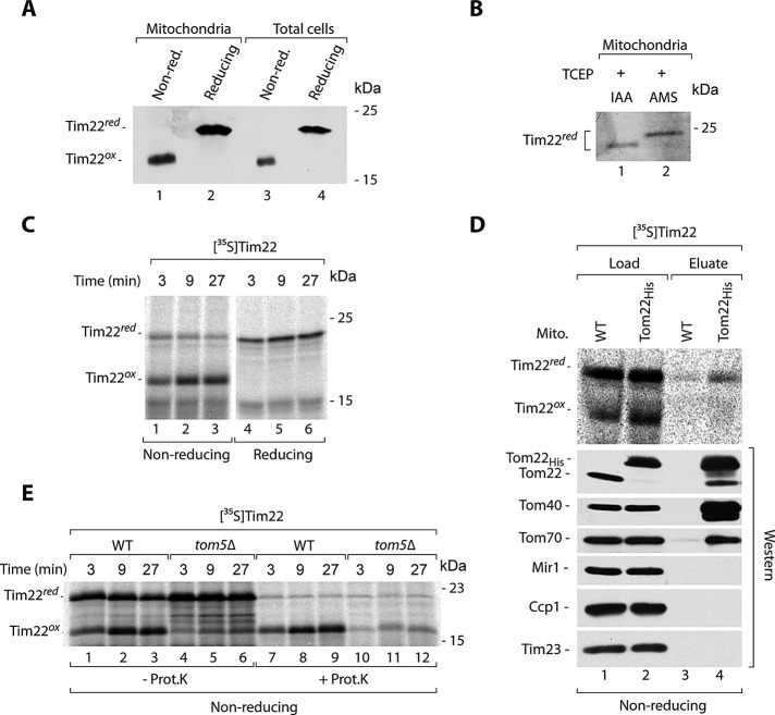
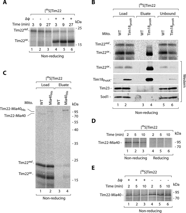
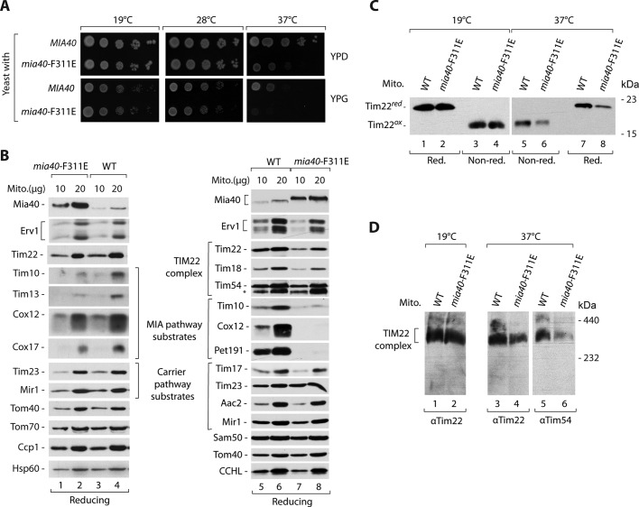
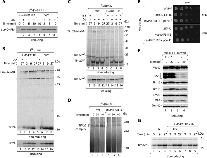
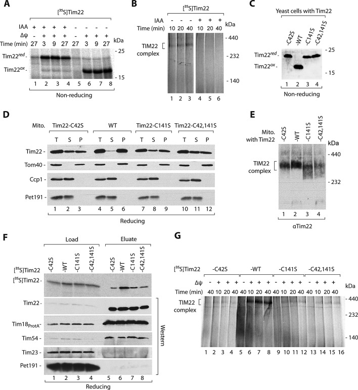
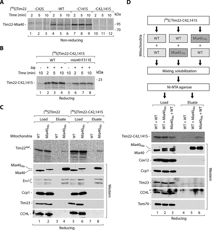
The mitochondrial intermembrane space assembly (MIA) pathway is generally considered to be dedicated to the redox-dependent import and biogenesis of proteins localized to the intermembrane space of mitochondria. The oxidoreductase Mia40 is a central component of the pathway responsible for the transfer of disulfide bonds to intermembrane space precursor proteins, causing their oxidative folding. Here we present the first evidence that the function of Mia40 is not restricted to the transport and oxidative folding of intermembrane space proteins. We identify Tim22, a multispanning membrane protein and core component of the TIM22 translocase of inner membrane, as a protein with cysteine residues undergoing oxidation during Tim22 biogenesis. We show that Mia40 is involved in the biogenesis and complex assembly of Tim22. Tim22 forms a disulfide-bonded intermediate with Mia40 upon import into mitochondria. Of interest, Mia40 binds the Tim22 precursor also via noncovalent interactions. We propose that Mia40 not only is responsible for disulfide bond formation, but also assists the Tim22 protein in its integration into the inner membrane of mitochondria.


Figures

FIGURE 1:
FIGURE 1:
Cysteine residues of Tim22 are present in the oxidized state. (A) Isolated wild-type mitochondria and total cell extracts were separated by SDS–PAGE, followed by immunodecoration with Tim22-specific antibodies. (B) Mitochondrial proteins were reduced with 25 mM TCEP, followed by incubation with 50 mM IAA or 15 mM AMS. The samples were separated by SDS–PAGE, followed by immunodecoration with Tim22-specific antibodies. (C) Radiolabeled Tim22 precursor protein was incubated with wild-type mitochondria. Nonimported protein was removed by proteinase K. (D) Radiolabeled Tim22 precursor protein was incubated for 5 min with mitochondria isolated from WT and Tom22His yeast strains. Mitochondria were solubilized in digitonin, and protein complexes were isolated via affinity purification. Load, 4%; eluate, 100%. (E) Radiolabeled Tim22 precursor was incubated with mitochondria isolated from WT and tom5Δ yeast strains. Where indicated, nonimported protein was removed by proteinase K. WT, wild type.
FIGURE 2:
FIGURE 2:
Tim22 interacts with Mia40. (A) Radiolabeled Tim22 precursor was imported into wild-type mitochondria in the presence or absence of the electrochemical IM potential (Δψ). Nonimported protein was removed by proteinase K. (B) Radiolabeled Tim22 precursor protein was incubated for 30 min with mitochondria isolated from WT and Tim18ProtA cells. Mitochondria were solubilized in digitonin, and protein complexes were isolated via affinity chromatography. Load, 3%; eluate, 100%; unbound 3%. (C) Mitochondria isolated from WT or Mia40His strains were incubated with radiolabeled Tim22 for 5 min. Mitochondria were solubilized in digitonin, and Mia40 conjugates were isolated by affinity purification. Load 3%; eluate 100%. (D) Radiolabeled Tim22 precursor was incubated with wild-type mitochondria and analyzed in the presence of IAA (nonreducing) or DTT (reducing). (E) Radiolabeled Tim22 precursor was incubated with wild-type mitochondria in the presence or absence of IM potential (Δψ), and Mia40 conjugates were analyzed. WT, wild type.
FIGURE 3:
FIGURE 3:
Mia40 promotes the TIM22 complex assembly. (A) Wild-type (MIA40) and mia40-F311E strains were subjected to consecutive 10-fold dilutions, spotted on YPD and YPG, and grown for 3–7 d at the indicated temperatures. (B, C) Mitochondria were isolated from WT and mia40-F311E mutant strains grown on YPG liquid medium at 19°C (lanes 1–4) or shifted to 37°C (lanes 5–8). (B) Mitochondrial proteins were analyzed by SDS–PAGE, followed by Western blotting. (C) The redox state of Tim22 was analyzed in the presence of IAA (nonreducing) or DTT (reducing). (D) Mitochondria were solubilized in digitonin-containing buffer, and the TIM22 complex was analyzed by blue native electrophoresis, followed by immunodecoration with Tim22- or Tim54-specific antibodies. WT, wild type.
FIGURE 4:
FIGURE 4:
Tim22 import is defective in the mia40-F311E mitochondria. (A–D) Radiolabeled precursors of Su9-DHFR, Tim9, and Tim22 were incubated with mitochondria isolated from WT and mia40-F311E mutant strains grown at 19°C. (A–C) Mitochondria were treated with proteinase K, except the Tim9 samples 1–8. (D) Mitochondria were solubilized in digitonin-containing buffer and analyzed by blue native electrophoresis. (E) Wild-type (MIA40) strain and mia40-F311E mutant transformed with the multicopy ERV1 plasmid were subjected to consecutive 10-fold dilutions, spotted on YPD and YPG, and grown for 2 d at 37°C. (F) Mitochondria were isolated from mia40-F311E– and mia40-F311E–overproducing Erv1 strain grown at 19°C and analyzed by Western blotting. (G) Radiolabeled Tim22 precursor was incubated with mitochondria isolated from the indicated strains. Mitochondria were treated with proteinase K. WT, wild type. Asterisk indicates a nonspecific band.
FIGURE 5:
FIGURE 5:
Cysteine residues of Tim22 are important for the Tim22 biogenesis. (A, B) Radiolabeled Tim22 precursor protein was preincubated for 5 min with or without 50 mM IAA. (A) Nonimported protein was removed by proteinase K. (B) Samples were solubilized in digitonin-containing buffer and analyzed by blue native electrophoresis. (C) Total protein extracts prepared from WT and tim22 cysteine mutant cells were separated on SDS–PAGE, followed by immunodecoration with Tim22-specific antibody. (D) Mitochondria isolated from WT and tim22 cysteine-residue mutants were subjected to alkaline carbonate extraction. T, total mitochondrial protein extract; S, supernatant; P, pellet of mitochondrial membranes. (E) Mitochondria isolated from WT and tim22 cysteine-residue mutants shifted to 37°C for 6 h were analyzed by blue native electrophoresis. (F) Radiolabeled wild-type Tim22 and Tim22 cysteine-mutant precursor proteins were incubated for 30 min with mitochondria isolated from Tim18ProtA yeast strains. Mitochondria were solubilized in digitonin-containing buffer, and protein complexes were isolated. Load, 3%; eluate, 100%. (G) Radiolabeled wild-type Tim22 and Tim22 cysteine-mutant precursor proteins were incubated with wild-type mitochondria. Mitochondria were solubilized in digitonin-containing buffer and analyzed by blue native electrophoresis. WT, wild type.
FIGURE 6:
FIGURE 6:
Mia40 binds the incoming Tim22 precursor in a covalent and noncovalent manner. (A) Radiolabeled wild-type Tim22 and Tim22 cysteine-mutant precursor proteins were incubated with wild-type mitochondria. (B) Radiolabeled Tim22-C42,141S precursor was incubated with the mitochondria isolated from WT and mia40-F311E mutant cells. Mitochondria were treated with proteinase K. (C) Mitochondria isolated from WT or Mia40His strains were incubated with radiolabeled Tim22 or Tim22-C42,141S for 5 min. Mitochondria were solubilized in digitonin buffer, and Mia40 complexes were isolated by affinity purification. Load, 3%; eluate, 100%. (D) Radiolabeled Tim22-C42,141S was incubated with WT or Mia40His mitochondria. Mitochondria were solubilized in digitonin with wild-type or Mia40His mitochondria as indicated and subjected to affinity purification. Load, 0,5%; eluate, 100%. (A–D) The samples were analyzed by digital autoradiography or Western blotting. WT, wild type. Asterisk indicates a nonspecific band.

Similar articles

Cited by

References

    1. Baker MJ, Webb CT, Stroud DA, Palmer CS, Frazier AE, Guiard B, Chacinska A, Gulbis JM, Ryan MT. Structural and functional requirements for activity of the Tim9-Tim10 complex in mitochondrial protein import. Mol Biol Cell. 2009;20:769–779. - PMC - PubMed
    1. Banci L, Bertini I, Calderone V, Cefaro C, Ciofi-Baffoni S, Gallo A, Kallergi E, Lionaki E, Pozidis C, Tokatlidis K. Molecular recognition and substrate mimicry drive the electron-transfer process between MIA40 and ALR. Proc Natl Acad Sci USA. 2011;108:4811–4816. - PMC - PubMed
    1. Banci L, et al. Molecular chaperone function of Mia40 triggers consecutive induced folding steps of the substrate in mitochondrial protein import. Proc Natl Acad Sci USA. 2010;107:20190–20195. - PMC - PubMed
    1. Banci L, Bertini I, Cefaro C, Ciofi-Baffoni S, Gallo A, Martinelli M, Sideris DP, Katrakili N, Tokatlidis K. MIA40 is an oxidoreductase that catalyzes oxidative protein folding in mitochondria. Nat Struct Mol Biol. 2009;16:198–206. - PubMed
    1. Becker T, Böttinger L, Pfanner N. Mitochondrial protein import: from transport pathways to an integrated network. Trends Biochem Sci. 2012;37:85–91. - PubMed

Publication types

MeSH terms

Substances

LinkOut - more resources