. 2013 Feb;24(4):474-82.
doi: 10.1091/mbc.E12-07-0521. Epub 2013 Jan 2.

αvβ8 integrin interacts with RhoGDI1 to regulate Rac1 and Cdc42 activation and drive glioblastoma cell invasion

Affiliations

αvβ8 integrin interacts with RhoGDI1 to regulate Rac1 and Cdc42 activation and drive glioblastoma cell invasion

Steve B Reyes et al. Mol Biol Cell. 2013 Feb.

Abstract

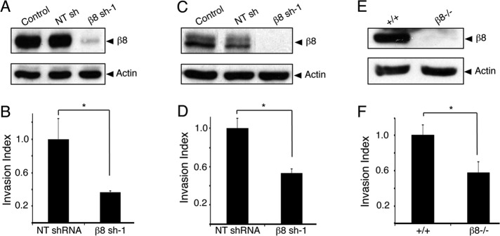
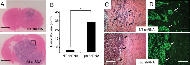
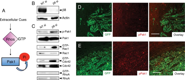
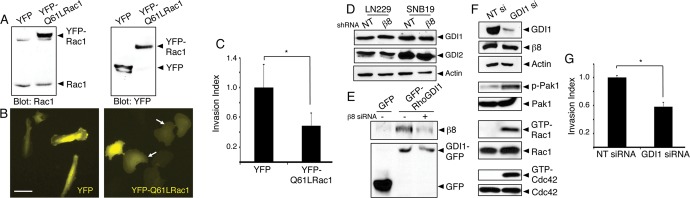
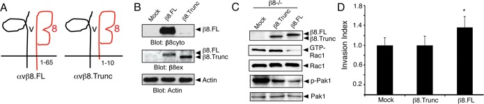
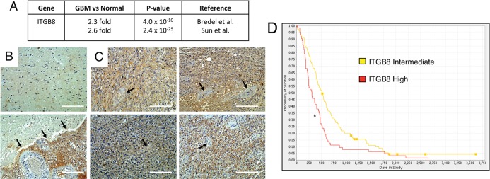
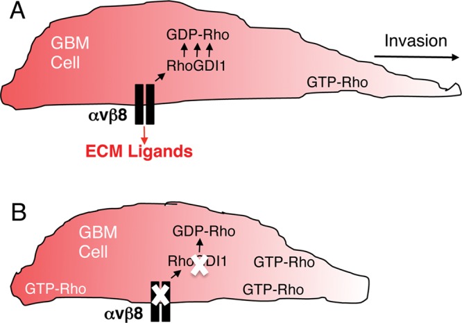
The malignant brain cancer glioblastoma multiforme (GBM) displays invasive growth behaviors that are regulated by extracellular cues within the neural microenvironment. The adhesion and signaling pathways that drive GBM cell invasion remain largely uncharacterized. Here we use human GBM cell lines, primary patient samples, and preclinical mouse models to demonstrate that integrin αvβ8 is a major driver of GBM cell invasion. β8 integrin is overexpressed in many human GBM cells, with higher integrin expression correlating with increased invasion and diminished patient survival. Silencing β8 integrin in human GBM cells leads to impaired tumor cell invasion due to hyperactivation of the Rho GTPases Rac1 and Cdc42. β8 integrin coimmunoprecipitates with Rho-GDP dissociation inhibitor 1 (RhoGDI1), an intracellular signaling effector that sequesters Rho GTPases in their inactive GDP-bound states. Silencing RhoGDI1 expression or uncoupling αvβ8 integrin-RhoGDI1 protein interactions blocks GBM cell invasion due to Rho GTPase hyperactivation. These data reveal for the first time that αvβ8 integrin, via interactions with RhoGDI1, regulates activation of Rho proteins to promote GBM cell invasiveness. Hence targeting the αvβ8 integrin-RhoGDI1 signaling axis might be an effective strategy for blocking GBM cell invasion.


Figures

FIGURE 1:
FIGURE 1:
β8 integrin drives GBM cell invasion in vitro. Human LN229 cells (A), SNB19 cells (C), and transformed mouse astrocytes (E) express robust levels of β8 integrin protein. Cells genetically null for β8 integrin or expressing β8 integrin shRNAs show diminished integrin protein expression. LN229 cells (B), SNB19 cells (D), and transformed mouse astrocytes (F) expressing diminished β8 integrin proteins display significantly reduced invasiveness; *p < 0.005.
FIGURE 2:
FIGURE 2:
β8 integrin is essential for GBM cell invasion in vivo. (A) Coronal sections from LN229 tumors expressing scrambled shRNAs (top) or β8 shRNAs (bottom) were analyzed by H&E staining. Mice (n = 6 per cell type) were injected with GBM cells. Representative images are shown. Scale bars, 2 mm. (B) Quantitation of tumor volumes, as determined by measuring cross-sectional areas of H&E–stained sections. LN229 cells expressing β8 shRNAs generate significantly larger tumors. Sections from six different tumors per cell type were analyzed; p < 0.001. (C) High magnification H&E–stained images from boxed areas in A. Coronal sections through LN229 tumors expressing scrambled shRNAs (top) or β8 shRNAs (bottom). Scale bars, 200 μm. (D) LN229 tumors expressing scrambled shRNAs (top) or β8 shRNAs (bottom) were immunofluorescently labeled with anti-GFP antibodies (green). Note that LN229 cells expressing scrambled shRNAs display robust patterns of perivascular invasion, whereas tumor cells expressing β8 shRNAs display diminished invasiveness. Scale bars, 100 μm.
FIGURE 3:
FIGURE 3:
β8 integrin suppresses Rac1 and Cdc42 activation in GBM cells. (A) Schematic model for regulation of Rho GTPase and Pak1 activation by extracellular cues. (B) Lysates from LN229 cells expressing scrambled siRNAs or siRNAs targeting β8 integrin were immunoblotted for β8 integrin or actin. (C) Integrin-dependent levels of phosphorylated-Pak1 or GTP-bound Rac1, Cdc42, and RhoA were analyzed in LN229 cell lysates. Phosphorylated Pak1 levels are increased after integrin silencing. Similarly, elevated levels of GTP-bound Rac1 and Cdc42, but not RhoA, were detected in cells expressing diminished levels of β8 integrin. (D, E) Intracranial tumors generated from LN229 cells expressing scrambled shRNAs (D) or β8 shRNAs (E) were analyzed by double immunofluorescence using anti–phospho-Pak1 (red) and anti-GFP (green) antibodies. At least three different sections from four different tumors were analyzed per cell type, with representative images shown. Note the elevated levels of phosphorylated Pak1 in tumor cells expressing β8 shRNAs. Scale bars, 100 μm.
FIGURE 4:
FIGURE 4:
Rac1 hyperactivation or RhoGDI1 inactivation leads to impaired GBM cell invasion. (A) Lysates from LN229 cells expressing YFP or YFP-tagged Q61L-Rac1 were immunoblotted with anti-Rac1 (left) or anti-GFP (right) antibodies, revealing similar levels of expression. (B) Images of LN229 cells expressing YFP (left) or YFP-tagged Q61L-Rac1 (right). Note the flattened morphologies of cells expressing Q61L-Rac1 (arrows). Scale bar, 60 μm. (C) Quantitation of three-dimensional invasive capacities of LN229 cells expressing YFP or YFP-Q61L-Rac1; *p < 0.05. (D) SNB19 and LN229 cells expressing scrambled shRNAs or β8 shRNAs were analyzed for endogenous RhoGDI1 and RhoGDI2 protein expression. Levels of RhoGDI proteins are not altered in cells expressing β8 shRNAs. (E) LN229 cells transfected with plasmids expressing GFP or GFP-tagged RhoGDI1 were lysed and immunoprecipitated with anti-GFP monoclonal antibodies and immunoblotted with anti-β8 integrin antibodies (top) or anti-GFP polyclonal antibodies (bottom). Note that β8 integrin and RhoGDI1 proteins coimmunoprecipitate, and these associations are reduced in LN229 cells expressing β8 siRNAs. (F) LN229 cells expressing scrambled siRNAs or siRNAs targeting RhoGDI1 were analyzed for RhoGDI1 protein expression. Silencing RhoGDI1 leads to elevated levels of phosphorylated Pak1 and increased levels of GTP-bound Rac1 and Cdc42. (G) Invasive behavior of LN229 cells expressing nontargeting siRNAs or RhoGD1 siRNAs quantified in invasion assays; *p < 0.05.
FIGURE 5:
FIGURE 5:
Uncoupling β8 integrin–RhoGDI1 interactions leads to enhanced Rac1 activation and diminished GBM cell invasion. (A) Diagram showing αvβ8.FL containing 65 amino acids in the β8 integrin cytoplasmic tail and αvβ8.Trunc containing only 10 amino acids in the β8 integrin tail. (B) β8.FL and β8.Trunc proteins were expressed in HEK-293T cells, and lysates were immunoblotted with antibodies recognizing the β8 integrin cytoplasmic (β8cyto) or extracellular domains (β8ex). (C) Expression of β8.FL in β8/ transformed astroglial progenitor cells reduces Rac1 activation, whereas levels of GTP-bound Rac1 remain high in cells expressing β8.Trunc protein. (D) In contrast to β8.FL, β8.Trunc protein does not ameliorate invasion defects in β8/-transformed cells; *p, **p = 0.05.
FIGURE 6:
FIGURE 6:
High ITGB8 expression in GBM correlates with reduced patient survival. (A) Oncomine summary of two independent transcriptome data sets comparing normal human brain samples to GBM samples. A greater-than-twofold increase in ITGB8 expression was detected in GBM samples as compared with normal brain samples. (B) Two different human brain sections containing neural tissue adjacent to the primary GBM were immunostained with anti–β8 integrin antibodies. Note the low levels of β8 integrin protein expression in the normal human brain. Bottom, a tumor margin revealing β8 integrin protein markedly upregulated in GBM cells. (C) Four different human GBM samples near the tumor core were immunolabeled with anti–β8 integrin antibodies. Note the robust levels of β8 integrin protein in tumor cells, with little if any integrin expression in intratumoral blood vessels (arrows). Scale bars, 200 μm. (D) Kaplan–Meier survival curve comparing human glioma samples with twofold or greater ITGB8 expression (n = 114) vs. samples with intermediate levels of ITGB8 expression (n = 225). Elevated ITGB8 expression correlates with diminished patient survival; *p < 0.001. Results were generated using the National Cancer Institute REMBRANDT database.
FIGURE 7:
FIGURE 7:
αvβ8 integrin–RhoGDI1 protein complexes regulate Rho GTPase activation and drive GBM cell invasion. (A) αvβ8 integrin is expressed in GBM cells, where it interacts with RhoGDI1 via the β8 cytoplasmic tail. αvβ8 integrin–RhoGDI1 protein complexes recruit GDP-bound Rac1 and Cdc42 to control activation of Rho proteins, thus driving GBM cell polarity and invasion. In addition, αvβ8 integrin likely binds to ECM protein ligands, including latent TGFβs, to regulate intracellular signaling pathways involved in GBM cell invasion. (B) Uncoupling αvβ8 integrin–RhoGDI1 protein complexes by mutating the integrin cytoplasmic tail or silencing β8 integrin or RhoGDI1 gene expression leads to elevated levels of GTP-bound Rho proteins, resulting in diminished GBM cell polarity and invasion.

Similar articles

Cited by

References

    1. Arias-Romero LE, Chernoff J. A tale of two Paks. Biol Cell. 2008;100:97–108. - PubMed
    1. Arnold TD, Ferrero GM, Qiu H, Phan IT, Akhurst RJ, Huang EJ, Reichardt LF. Defective retinal vascular endothelial cell development as a consequence of impaired integrin alphavbeta8-mediated activation of transforming growth factor-beta. J Neurosci. 2012;32:1197–1206. - PMC - PubMed
    1. Batchelor TT, et al. AZD2171, a pan-VEGF receptor tyrosine kinase inhibitor, normalizes tumor vasculature and alleviates edema in glioblastoma patients. Cancer Cell. 2007;11:83–95. - PMC - PubMed
    1. Boulter E, Garcia-Mata R, Guilluy C, Dubash A, Rossi G, Brennwald PJ, Burridge K. Regulation of Rho GTPase crosstalk, degradation and activity by RhoGDI1. Nat Cell Biol. 2010;12:477–483. - PMC - PubMed
    1. Bredel M, Bredel C, Juric D, Harsh GR, Vogel H, Recht LD, Sikic BI. High-resolution genome-wide mapping of genetic alterations in human glial brain tumors. Cancer Res. 2005;65:4088–4096. - PubMed

Publication types

MeSH terms