Review
. 2013 Jan 1;5(1):a008334.
doi: 10.1101/cshperspect.a008334.

Development of the endochondral skeleton

Affiliations
Review

Development of the endochondral skeleton

Fanxin Long et al. Cold Spring Harb Perspect Biol. .

Abstract

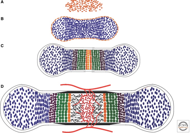
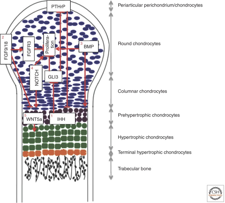
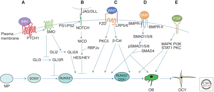
Much of the mammalian skeleton is composed of bones that originate from cartilage templates through endochondral ossification. Elucidating the mechanisms that control endochondral bone development is critical for understanding human skeletal diseases, injury response, and aging. Mouse genetic studies in the past 15 years have provided unprecedented insights about molecules regulating chondrocyte formation, chondrocyte maturation, and osteoblast differentiation, all key processes of endochondral bone development. These include the roles of the secreted proteins IHH, PTHrP, BMPs, WNTs, and FGFs, their receptors, and transcription factors such as SOX9, RUNX2, and OSX, in regulating chondrocyte and osteoblast biology. This review aims to integrate the known functions of extracellular signals and transcription factors that regulate development of the endochondral skeleton.


Figures

Figure 1.
Figure 1.
Endochondral bone development. (A) Mesenchymal condensation. (B) Chondrocyte differentiation. Cells centrally located within the mesenchymal condensation differentiate to chondrocytes (blue), whereas the peripheral cells form the perichondrium (gold). (C) Chondrocyte maturation. After the initial proliferation, chondrocytes at the center of the cartilage primordium undergo progressive maturation through prehypertrophy (brown), hypertrophy (green), and terminal hypertrophy (orange). (D) Cartilage vascularization and bone collar formation. Following terminal hypertrophy of chondrocytes, blood vessels (red lines) from the surrounding tissue invade the center of the hypertrophic zone, concurrent with formation of the bone collar (black) from the surrounding perichondrium. Vascular invasion leads to resorption of cartilage matrix, formation of the marrow (red), and deposition of bone (black) within the marrow cavity.
Figure 2.
Figure 2.
Extracellular signals regulating growth plate development. Depicted is a longitudinal section through one of two growth plates of a mouse long bone during late embryogenesis (E15.5–E19). The growth plate at this stage is without a secondary ossification center and is organized into distinct domains as indicated. (1) IHH and PTHrP coordinate chondrocyte proliferation and maturation through a negative-feedback mechanism. IHH produced by pre- and early hypertrophic chondrocytes stimulates chondrocyte proliferation and PTHrP transcription through derepression of GLI3. PTHrP in turn suppresses chondrocyte maturation associated with IHH expression. Direct IHH signaling also regulates the formation of columnar chondrocytes from round chondrocytes (not depicted here). (2) FGF9/18 from the perichondrium suppresses chondrocyte proliferation and maturation. FGFR3 expressed in chondrocytes is a likely receptor for FGF9/18 to suppress proliferation in the growth plate late in embryonic development and during postnatal bone growth. FGF9/18 may use other yet-to-be-established mechanisms to suppress chondrocyte maturation in the early embryo. (3) BMPs expressed by both chondrocytes and perichondrial cells promote proliferation and maturation. (4) NOTCH signaling in chondrocytes promotes proliferation and maturation. (5) WNT5A expressed by prehypertrophic chondrocytes stimulates hypertrophy.
Figure 3.
Figure 3.
Extracellular signals regulating osteoblast differentiation. Model is based on studies of the mouse limb skeleton. Osteoblasts differentiate from mesenchymal progenitors (MP) through distinct developmental stages marked by expression of key transcription factors including SOX9, RUNX2, and OSX. Mature osteoblasts (OB) can further differentiate to osteocytes (OCY) or bone lining cells (not depicted) or undergo apoptosis (not depicted). (A) Indian hedgehog (IHH) signaling is required for osteoblast differentiation during endochondral bone development. IHH binding to the receptor Patched homolog 1 (PTCH1) activates signaling through Smoothened (SMO), thereby inhibiting the generation of the proteolytically cleaved GLI3 repressor (GLI3R) and promoting the generation of the full-length GLI2 activator (GLI2A). Whereas derepression of GLI3R is sufficient to generate RUNX2+ cells, both derepression of GLI3R and activation of GLI2A are necessary for progression to the RUNX2+OSX+ stage. (B) NOTCH signaling inhibits osteoblast differentiation. Following binding to their ligands, Jagged (JAG) or Delta-like (DLL), Notch receptors are proteolytically cleaved by the γ-secretase complex, leading to release of the Notch intracellular domain (NICD) from the plasma membrane. NICD interacts with RBPJκ and together they activate downstream target genes, including HES (Hairy and Enhancer of Split) and HEY (HES-related with YRPW motif) family transcription factors, ultimately leading to inhibition of osteoblast differentiation, seemingly at a stage before OSX activation. (C) WNT signaling promotes osteoblast differentiation. During β-catenin-dependent WNT signaling, β-catenin is stabilized following binding of WNT to its receptors Frizzled (FZD) and lipoprotein receptor-related protein 5 (LRP5) or LRP6, leading to the transcription of β-catenin target genes and ultimately stimulating progression from the RUNX2+ stage to the RUNX2+OSX+ stage, and from RUNX2+OSX+ cells to mature osteoblasts. WNT can also signal independently of LRP5/6 and β-catenin through protein kinase Cδ (PKCδ), promoting progression to the RUNX2+OSX+ stage through an unknown mechanism. (D) Bone morphogenetic protein (BMP) signaling stimulates osteoblast differentiation and function. Binding of BMP2 or BMP4 to their receptors results in phosphorylation of SMAD1, SMAD5, or SMAD8. These can then form a complex with their partner, SMAD4, and enter the nucleus to regulate gene expression, ultimately promoting the transition to RUNX2+OSX+ cells and enhancing the function of mature osteoblasts; however, a direct role for SMAD signaling in osteoblast differentiation remains to be shown. (E) Fibroblast growth factor (FGF) signaling has diverse roles in osteoblast lineage cells. FGFs function by binding to cell surface Tyr kinase FGF receptors (FGFR1–FGFR4 in humans and mice), leading to the activation of multiple signaling modules. FGF signaling regulates preosteoblast proliferation and osteoblast differentiation, as well as the function of mature osteoblasts. However, the precise stages at which FGFs regulate proliferation and differentiation, and the intracellular signaling cascades responsible for each function, remain to be elucidated. BMPR, BMP receptor; MAPK, mitogen-activated protein kinase; PI3K, phosphoinositide 3-kinase; STAT1, signal transducer and activator of transcription 1.

References

    1. Ai M, Holmen SL, Van Hul W, Williams BO, Warman ML 2005. Reduced affinity to and inhibition by DKK1 form a common mechanism by which high bone mass-associated missense mutations in LRP5 affect canonical Wnt signaling. Mol Cell Biol 25: 4946–4955 - PMC - PubMed
    1. Akiyama H, Chaboissier MC, Martin JF, Schedl A, de Crombrugghe B 2002. The transcription factor Sox9 has essential roles in successive steps of the chondrocyte differentiation pathway and is required for expression of Sox5 and Sox6. Genes Dev 16: 2813–2828 - PMC - PubMed
    1. Akiyama H, Lyons JP, Mori-Akiyama Y, Yang X, Zhang R, Zhang Z, Deng JM, Taketo MM, Nakamura T, Behringer RR, et al. 2004. Interactions between Sox9 and β-catenin control chondrocyte differentiation. Genes Dev 18: 1072–1087 - PMC - PubMed
    1. Akiyama H, Kim JE, Nakashima K, Balmes G, Iwai N, Deng JM, Zhang Z, Martin JF, Behringer RR, Nakamura T, et al. 2005. Osteo-chondroprogenitor cells are derived from Sox9 expressing precursors. Proc Natl Acad Sci 102: 14665–14670 - PMC - PubMed
    1. Albers J, Schulze J, Beil FT, Gebauer M, Baranowsky A, Keller J, Marshall RP, Wintges K, Friedrich FW, Priemel M, et al. 2011. Control of bone formation by the serpentine receptor Frizzled-9. J Cell Biol 192: 1057–1072 - PMC - PubMed

Publication types

Substances