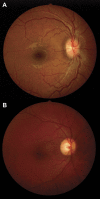
Nonmydriatic ocular fundus photography among headache patients in an emergency department
- PMID: 23284060
- PMCID: PMC3590046
- DOI: 10.1212/WNL.0b013e31827f0f20
Nonmydriatic ocular fundus photography among headache patients in an emergency department
Abstract
Objectives: Determine the frequency of and the predictive factors for abnormal ocular fundus findings among emergency department (ED) headache patients.
Methods: Cross-sectional study of prospectively enrolled adult patients presenting to our ED with a chief complaint of headache. Ocular fundus photographs were obtained using a nonmydriatic fundus camera that does not require pupillary dilation. Demographic and neuroimaging information was collected. Photographs were reviewed independently by 2 neuroophthalmologists for findings relevant to acute care. The results were analyzed using univariate statistics and logistic regression modeling.
Results: We included 497 patients (median age: 40 years, 73% women), among whom 42 (8.5%, 95% confidence interval: 6%-11%) had ocular fundus abnormalities. Of these 42 patients, 12 had disc edema, 9 had optic nerve pallor, 6 had grade III/IV hypertensive retinopathy, and 15 had isolated retinal hemorrhages. Body mass index ≥ 35 kg/m(2) (odds ratio [OR]: 2.3, p = 0.02), younger age (OR: 0.7 per 10-year increase, p = 0.02), and higher mean arterial blood pressure (OR: 1.3 per 10-mm Hg increase, p = 0.003) were predictive of abnormal retinal photography. Patients with an abnormal fundus had a higher percentage of hospital admission (21% vs 10%, p = 0.04). Among the 34 patients with abnormal ocular fundi who had brain imaging, 14 (41%) had normal imaging.
Conclusions: Ocular fundus abnormalities were found in 8.5% of patients with headache presenting to our ED. Predictors of abnormal funduscopic findings included higher body mass index, younger age, and higher blood pressure. Our study confirms the importance of funduscopic examination in patients with headache, particularly in the ED, and reaffirms the utility of nonmydriatic fundus photography in this setting.
Figures

Comment in
-
Nonmydriatic ocular fundus photography among headache patients in an emergency department.Neurology. 2013 Aug 20;81(8):774-5. doi: 10.1212/WNL.0b013e3182a293e1. Neurology. 2013. PMID: 23960195 No abstract available.
-
Nonmydriatic ocular fundus photography among headache patients in an emergency department.Neurology. 2013 Oct 8;81(15):1366. doi: 10.1212/01.wnl.0000436284.64959.67. Neurology. 2013. PMID: 24101750 No abstract available.
-
Reply: To PMID 23284060.Neurology. 2013 Oct 8;81(15):1366-7. Neurology. 2013. PMID: 24244961 No abstract available.
References
-
- Pitts SR, Niska RW, Xu J, Burt CW. National Hospital Ambulatory Medical Care Survey: 2006 emergency department summary. Natl Health Stat Report 2008;(7):1–38 - PubMed
-
- Lucado J, Paez K, Elixhauser A. Headaches in U.S. Hospitals and Emergency Departments, 2008: HCUP Statistical Brief #111. Rockville: Agency for Healthcare Research and Quality; 2011 - PubMed
-
- Linet MS, Stewart WF, Celentano DD, Ziegler D, Sprecher M. An epidemiologic study of headache among adolescents and young adults. JAMA 1989;261:2211–2216 - PubMed
-
- Goldstein JN, Camargo CA, Jr, Pelletier AJ, Edlow JA. Headache in United States emergency departments: demographics, work-up and frequency of pathological diagnoses. Cephalalgia 2006;26:684–690 - PubMed
-
- Olesen J. The international classification of headache disorders. 2nd edition (ICHD-II). Rev Neurol (Paris) 2005;161:689–691 - PubMed
Publication types
MeSH terms
Grants and funding
LinkOut - more resources
Full Text Sources
Other Literature Sources
Medical
