. 2013 Jan 15;110(3):912-7.
doi: 10.1073/pnas.1220895110. Epub 2013 Jan 2.

RAC1P29S is a spontaneously activating cancer-associated GTPase

Affiliations

RAC1P29S is a spontaneously activating cancer-associated GTPase

Matthew J Davis et al. Proc Natl Acad Sci U S A. .

Abstract

RAC1 is a small, Ras-related GTPase that was recently reported to harbor a recurrent UV-induced signature mutation in melanoma, resulting in substitution of P29 to serine (RAC1(P29S)), ranking this the third most frequently occurring gain-of-function mutation in melanoma. Although the Ras family GTPases are mutated in about 30% of all cancers, mutations in the Rho family GTPases have rarely been observed. In this study, we demonstrate that unlike oncogenic Ras proteins, which are primarily activated by mutations that eliminate GTPase activity, the activated melanoma RAC1(P29S) protein maintains intrinsic GTP hydrolysis and is spontaneously activated by substantially increased inherent GDP/GTP nucleotide exchange. Determination and comparison of crystal structures for activated RAC1 GTPases suggest that RAC1(F28L)--a known spontaneously activated RAC1 mutant--and RAC1(P29S) are self-activated in distinct fashions. Moreover, the mechanism of RAC1(P29S) and RAC1(F28L) activation differs from the common oncogenic mutations found in Ras-like GTPases that abrogate GTP hydrolysis. The melanoma RAC1(P29S) gain-of-function point mutation therefore represents a previously undescribed class of cancer-related GTPase activity.


Conflict of interest statement

The authors declare no conflict of interest.

Figures

Fig. 1.
Fig. 1.
Structural map of RAC1. Structure of GMP-PNP–loaded RAC1WT (PDB ID code 3TH5) (12) with key regions indicated shown in cartoon format. Red spheres identify the residues described in the paper. The exploded view shows the Switch regions with side chains in cylinder format. Structural figures generated using CCP4MG (15).
Fig. 2.
Fig. 2.
In vitro nucleotide exchange assays. (A) Nucleotide exchange comparison of RAC1WT and RAC1P29S. RAC1P29S is a self-activating mutant as indicated by inherent self-association of mGTPγS without the addition of EDTA (Left). EDTA (20 mM) is added to facilitate RAC1WT nucleotide exchange (Right). In both experimental conditions, RAC1WT is significantly slower than RAC1P29S (P < 0.05). (B) Nucleotide exchange rate. Comparison of RAC1WT, RAC1P29S, and RAC1F28L with (Right) or without (Left) 20 mM EDTA addition. RAC1WT displays marginal exchange without addition of EDTA. SEM for three experiments shown. (C) Orthogonal nucleotide exchange pull-down assay. In vitro pull-down of activated GTPγS-loaded RAC1 constructs with the GTPase-binding domain of PAK1. Robust pull-down is observed for RAC1P29S and RAC1F28L with and without 10 mM EDTA. RAC1WT requires 10 mM EDTA to pull-down robustly, and dominant-negative RAC1T17N cannot load GTPγS. Loading controls shown in Fig. S2 A and B.
Fig. 3.
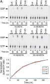
Fig. 3.
In vitro GTP hydrolysis assays. (A) EDTA tuning of the RAC1 hydrolysis assay. TLC separation of hydrolyzed [α-32P]GTP to [α-32P]GDP is shown for RAC1WT, RAC1P29S, RAC1F28L, and RAC1Q61L under 0, 5, 10, and 20 mM EDTA conditions in a reaction solution containing 10 mM MgCl2. (B) Time course of RAC1 first-order GTP hydrolysis assay. Quantification (GDP/GTP ratio) of TLC separation of hydrolyzed [α-32P]GTP to [α-32P]GDP for RAC1WT, RAC1P29S, RAC1F28L, and RAC1Q61L taken at 0, 30, 60, and 120 min. SEM for three experiments <0.1. Fig. S3 contains the SEM errors and reactions for each RAC1 GTPase analyzed.
Fig. 4.
Fig. 4.
Comparison of GTP analog-loaded RAC1 crystal structures. Crystal structures of RAC1F28L and RAC1Q61L are compared with RAC1WT (PDB ID code 3TH5) (12) and RAC1P29S (PDB ID code 3SBD) (12). Dashed box indicates region shown in the exploded views. Residues mutated in this study are indicated.
Fig. 5.
Fig. 5.
RAC1P29S exhibits an activated cellular phenotype in live and fixed cells. (A) Live COS-7 cells ectopically expressing GFP-RAC1WT, GFP-RAC1P29S, GFP-RAC1F28L, and GFP-RAC1Q61L were imaged using spinning-disk confocal microscopy. Membrane ruffles as indicated by accumulation of GFP-RAC1 signal were observed for GFP-RAC1P29S–, GFP-RAC1F28L–, and GFP-RAC1Q61L–expressing cells. RAC1 WT exhibited a more diffuse pattern with marginal membrane ruffling at cell edges. (B) NIH 3T3 cells stably expressing 3×-Flag-RAC1WT, 3×-Flag-RAC1P29S, and 3×-Flag-RAC1F28L were immunostained with anti-FLAG and probed with Alexa-Fluor 488 to visualize RAC1 expression, and subcellular localization and integrity of F-actin–containing stress-fibers was visualized through staining with rhodamine phalloidin. In both RAC1P29S- and RAC1F28L-expressing cells, membrane ruffling was observed as in COS-7 cells; ruffling is indicated with white arrows.

References

    1. Vetter IR, Wittinghofer A. The guanine nucleotide-binding switch in three dimensions. Science. 2001;294(5545):1299–1304. - PubMed
    1. Jaffe AB, Hall A. Rho GTPases: Biochemistry and biology. Annu Rev Cell Dev Biol. 2005;21:247–269. - PubMed
    1. Vega FM, Ridley AJ. Rho GTPases in cancer cell biology. FEBS Lett. 2008;582(14):2093–2101. - PubMed
    1. Ellenbroek SI, Collard JG. Rho GTPases: Functions and association with cancer. Clin Exp Metastasis. 2007;24(8):657–672. - PubMed
    1. Fritz G, Just I, Kaina B. Rho GTPases are over-expressed in human tumors. Int J Cancer. 1999;81(5):682–687. - PubMed

Publication types

MeSH terms