

CSF and brain structural imaging markers of the Alzheimer's pathological cascade
- PMID: 23284610
- PMCID: PMC3526616
- DOI: 10.1371/journal.pone.0047406
CSF and brain structural imaging markers of the Alzheimer's pathological cascade
Abstract
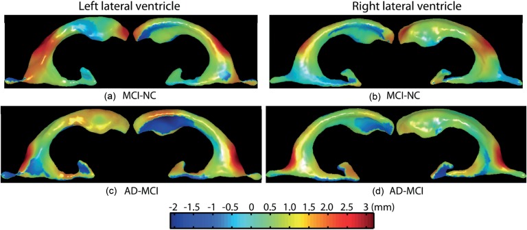
Cerebral spinal fluid (CSF) and structural imaging markers are suggested as biomarkers amended to existing diagnostic criteria of mild cognitive impairment (MCI) and Alzheimer's disease (AD). But there is no clear instruction on which markers should be used at which stage of dementia. This study aimed to first investigate associations of the CSF markers as well as volumes and shapes of the hippocampus and lateral ventricles with MCI and AD at the baseline and secondly apply these baseline markers to predict MCI conversion in a two-year time using the Alzheimer's Disease Neuroimaging Initiative (ADNI) cohort. Our results suggested that the CSF markers, including Aβ42, t-tau, and p-tau, distinguished MCI or AD from NC, while the Aβ42 CSF marker contributed to the differentiation between MCI and AD. The hippocampal shapes performed better than the hippocampal volumes in classifying NC and MCI, NC and AD, as well as MCI and AD. Interestingly, the ventricular volumes were better than the ventricular shapes to distinguish MCI or AD from NC, while the ventricular shapes showed better accuracy than the ventricular volumes in classifying MCI and AD. As the CSF markers and the structural markers are complementary, the combination of them showed great improvements in the classification accuracies of MCI and AD. Moreover, the combination of these markers showed high sensitivity but low specificity for predicting conversion from MCI to AD in two years. Hence, it is feasible to employ a cross-sectional sample to investigate dynamic associations of the CSF and imaging markers with MCI and AD and to predict future MCI conversion. In particular, the volumetric information may be good for the early stage of AD, while morphological shapes should be considered as markers in the prediction of MCI conversion to AD together with the CSF markers.
Conflict of interest statement
Figures


References
-
- McKhann GM, Knopman DS, Chertkow H, Hyman BT, Jack CR Jr, et al. (2011) The diagnosis of dementia due to Alzheimer's disease: recommendations from the National Institute on Aging-Alzheimer's Association workgroups on diagnostic guidelines for Alzheimer's disease. Alzheimers Dement 7: 263–269. - PMC - PubMed
-
- Albert MS, DeKosky ST, Dickson D, Dubois B, Feldman HH, et al. (2011) The diagnosis of mild cognitive impairment due to Alzheimer's disease: recommendations from the National Institute on Aging-Alzheimer's Association workgroups on diagnostic guidelines for Alzheimer's disease. Alzheimers Dement 7: 270–279. - PMC - PubMed
-
- Sperling RA, Aisen PS, Beckett LA, Bennett DA, Craft S, et al. (2011) Toward defining the preclinical stages of Alzheimer's disease: recommendations from the National Institute on Aging-Alzheimer's Association workgroups on diagnostic guidelines for Alzheimer's disease. Alzheimers Dement 7: 280–292. - PMC - PubMed
-
- Ferrarini L, Palm WM, Olofsen H, van der Landen R, van Buchem MA, et al. (2008) Ventricular shape biomarkers for Alzheimer's disease in clinical MR images. Magnetic resonance in medicine : official journal of the Society of Magnetic Resonance in Medicine/Society of Magnetic Resonance in Medicine 59: 260–267. - PubMed
Publication types
MeSH terms
Substances
LinkOut - more resources
Full Text Sources
Medical
Research Materials
