. 2012;7(12):e51223.
doi: 10.1371/journal.pone.0051223. Epub 2012 Dec 20.

Resveratrol attenuates ischemia/reperfusion injury in neonatal cardiomyocytes and its underlying mechanism

Affiliations

Resveratrol attenuates ischemia/reperfusion injury in neonatal cardiomyocytes and its underlying mechanism

Min Shen et al. PLoS One. 2012.

Abstract

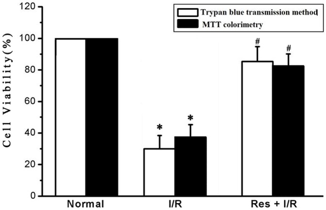
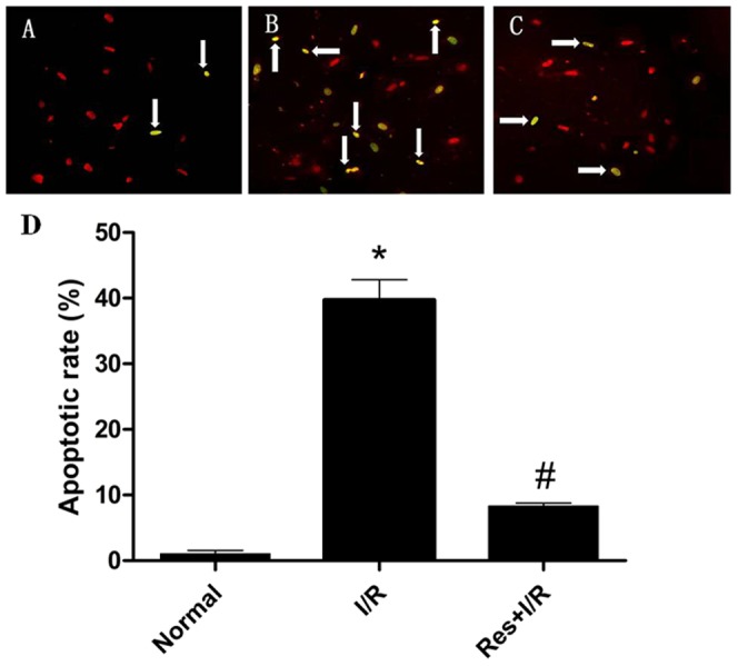
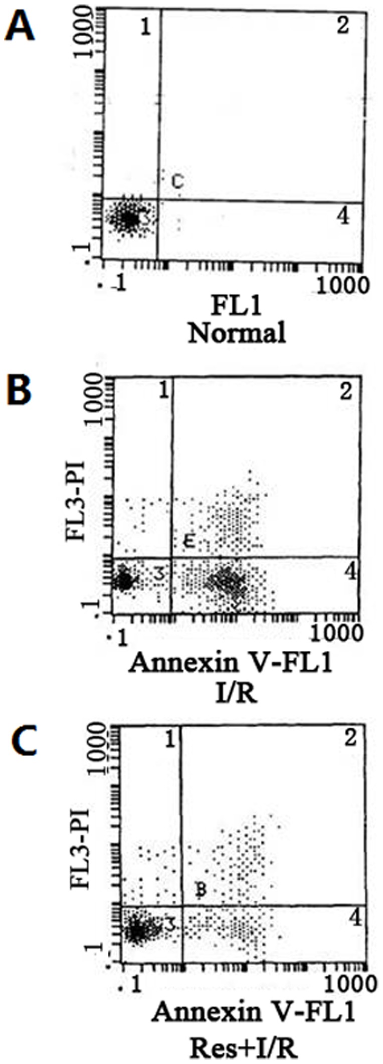
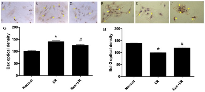
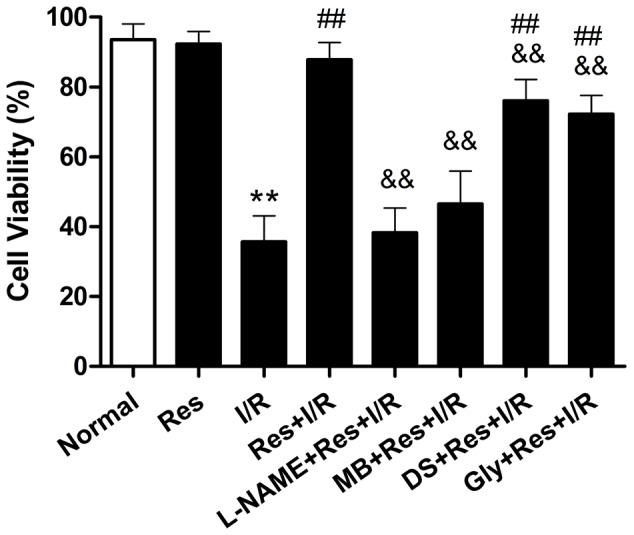
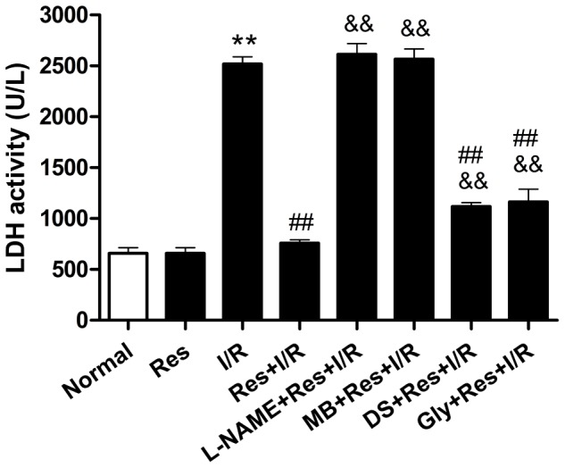
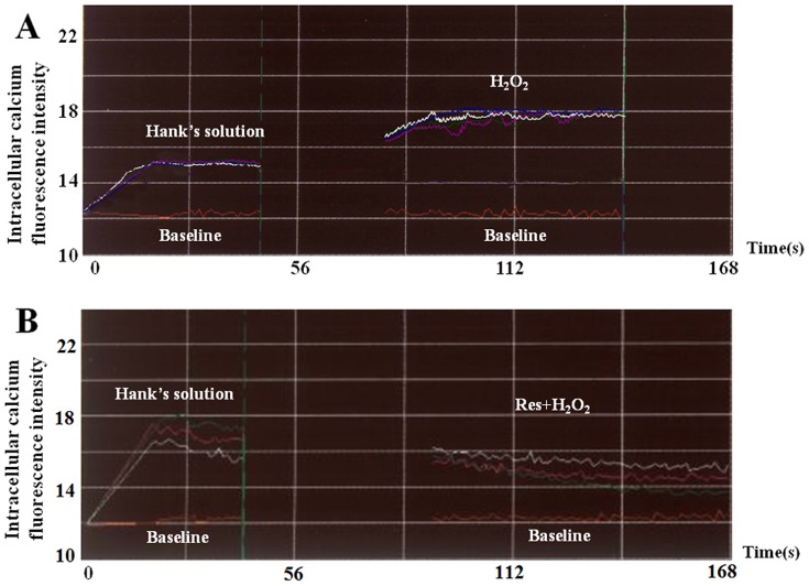
This study was designed to investigate whether Resveratrol (Res) could be a prophylactic factor in the prevention of I/R injury and to shed light on its underlying mechanism. Primary culture of neonatal rat cardiomyocytes were randomly distributed into three groups: the normal group (cultured cardiomyocytes were in normal conditions), the I/R group (cultured cardiomyocytes were subjected to 2 h simulated ischemia followed by 4 h reperfusion), and the Res+I/R group (100 µmol/L Res was administered before cardiomyocytes were subjected to 2 h simulated ischemia followed by 4 h reperfusion). To test the extent of cardiomyocyte injury, several indices were detected including cell viability, LDH activity, Na(+)-K(+)-ATPase and Ca(2+)-ATPase activity. To test apoptotic cell death, caspase-3 activity and the expression of Bcl-2/Bax were detected. To explore the underlying mechanism, several inhibitors, intracellular calcium, SOD activity and MDA content were used to identify some key molecules involved. Res increased cell viability, Na(+)-K(+)-ATPase and Ca(2+)-ATPase activity, Bcl-2 expression, and SOD level. While LDH activity, capase-3 activity, Bax expression, intracellular calcium and MDA content were decreased by Res. And the effect of Res was blocked completely by either L-NAME (an eNOS inhibitor) or MB (a cGMP inhibitor), and partly by either DS (a PKC inhibitor) or Glybenclamide (a K(ATP) inhibitor). Our results suggest that Res attenuates I/R injury in cardiomyocytes by preventing cell apoptosis, decreasing LDH release and increasing ATPase activity. NO, cGMP, PKC and K(ATP) may play an important role in the protective role of Res. Moreover, Res enhances the capacity of anti-oxygen free radical and alleviates intracellular calcium overload in cardiomyocytes.


Conflict of interest statement

Competing Interests: The authors have declared that no competing interests exist.

Figures

Figure 1
Figure 1. Effect of Res on cell viability by trypan blue method and methyl thiazolyl tetrazolium (MTT) colorimetry.
The two methods revealed that Res stabilized cell membrane and increase cell viability during I/R. Res, resveratrol; I/R, ischemia/reperfusion. *P<0.01 versus normal group, #P<0.01 versus I/R group. Values are the means ± SD, n = 10 in each group.
Figure 2
Figure 2. TUNEL staining and the apoptotic rate of cardiomyocytes.
The arrow refers to apoptotic cells. (A) In the normal group, a very few apoptotic cells were found. (B) In the I/R group, the apoptotic cells increased dramatically. (C) In the Res+I/R group, the number of apoptotic cells was far less than that in the I/R group, which suggested that Res lowered cell apoptosis induced by I/R. (D) The statistical data of apoptotic rate. *P<0.01 versus normal group, #P<0.01 versus I/R group. Values are the means ± SD, n = 10 in each group.
Figure 3
Figure 3. Assay of cardiomyocytic apoptosis with flow cytometry.
(A) In the normal group, most of the cells lied in left lower quadrant, which indicated that the cells were viable. (B) In the I/R group, the number of cells in the right lower quadrant increased obviously compared with that in (A), which suggested that I/R injury induced apoptosis. (C) In the Res+I/R group, the number of cells in the right lower quadrant decreased dramatically compared with that in (B), which suggested that Res lowered cell apoptosis triggered by I/R.
Figure 4
Figure 4. The expression of Bax and Bcl-2 detected by immunohistochemistry.
The expression of Bax and Bcl-2 were shown and arrows refer to them. (A)(D)In normal condition, Bax and Bcl-2 were expressed in cardiomyocytes. (B)(E) I/R injury promoted Bax expression and suppressed Bcl-2 expression. (C)(F)On the contrary, Res suppressed Bax expression and promoted Bcl-2 expression. (G) The statistical data of Bax optical density. (H) The statistical data of Bcl-2 optical density. *P<0.01 versus normal group, #P<0.01 versus I/R group. Values are the means ± SD, n = 10 in each group.
Figure 5
Figure 5. Effect of L-NAME, MB, DS and Gly on the cardiomyocytes viability in Res+I/R group.
Res protected the cell viability against I/R, the role of Res on the cell viability was almost abolished by L-NAME and MB, and was significantly attenuated by DS and Gly. L-NAME, a nitric oxide synthase inhibitor; MB, Methylene blue, a cGMP inhibitor; DS, D-Sphingosine, a PKC inhibitor; Gly, Glibenclamide, a KATP blocker. ** P<0.01 versus the normal group. ## P<0.01 versus the I/R group. && P<0.01 versus the Res+I/R group. Values are the means ± SD, n = 10 in each group.
Figure 6
Figure 6. Effect of L-NAME, MB, DS and Gly on the LDH activity in Res+I/R group.
The LDH activity in normal group was 658.1±97.2, while that in I/R group was 2517.9±124.5 and in Res+I/R group 758.8±54.6, which had a significant difference compared with I/R group. The effect of Res on the LDH activity was abolished by L-NAME and MB, and was significantly attenuated by DS and Gly. L-NAME, a nitric oxide synthase inhibitor; MB, Methylene blue, a cGMP inhibitor; DS, D-Sphingosine, a PKC inhibitor; Gly, Glibenclamide, a KATP blocker. ** P<0.01 versus the normal group. ## P<0.05 versus the I/R group. && P<0.05 versus the Res+I/R group. Values are the means ± SD, n = 10 in each group.
Figure 7
Figure 7. Effect of Res on the elevated intracellular calcium induced by H2O2.
(A) In Hank's solution, H2O2 led to the increase of intracellular calcium, and the fluorescence intensity increased. (B) In Res group, the effect of H2O2 was abolished by Res.

References

    1. Yellon DM, Hausenloy DJ (2007) Myocardial reperfusion injury. N Engl J Med 357: 1121–1135. - PubMed
    1. Chen Z, Chua CC, Ho YS, Hamdy RC, Chua BH (2001) Overexpression of Bcl-2 attenuates apoptosis and protects against myocardial I/R injury in transgenic mice. Am J Physiol Heart Circ Physiol 280: H2313–2320. - PubMed
    1. Kajstura J, Cheng W, Reiss K, Clark WA, Sonnenblick EH, et al. (1996) Apoptotic and necrotic myocyte cell deaths are independent contributing variables of infarct size in rats. Lab Invest 74: 86–107. - PubMed
    1. Turer AT, Hill JA (2010) Pathogenesis of myocardial ischemia-reperfusion injury and rationale for therapy. Am J Cardiol 106: 360–368. - PMC - PubMed
    1. Gross A, McDonnell JM, Korsmeyer SJ (1999) BCL-2 family members and the mitochondria in apoptosis. Genes Dev 13: 1899–1911. - PubMed

Publication types

MeSH terms