. 2012;7(12):e51672.
doi: 10.1371/journal.pone.0051672. Epub 2012 Dec 20.

Heterologous SUMO-2/3-ubiquitin chains optimize IκBα degradation and NF-κB activity

Affiliations

Heterologous SUMO-2/3-ubiquitin chains optimize IκBα degradation and NF-κB activity

Fabienne Aillet et al. PLoS One. 2012.

Abstract

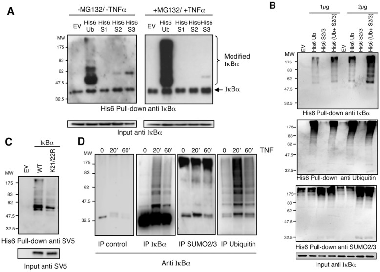
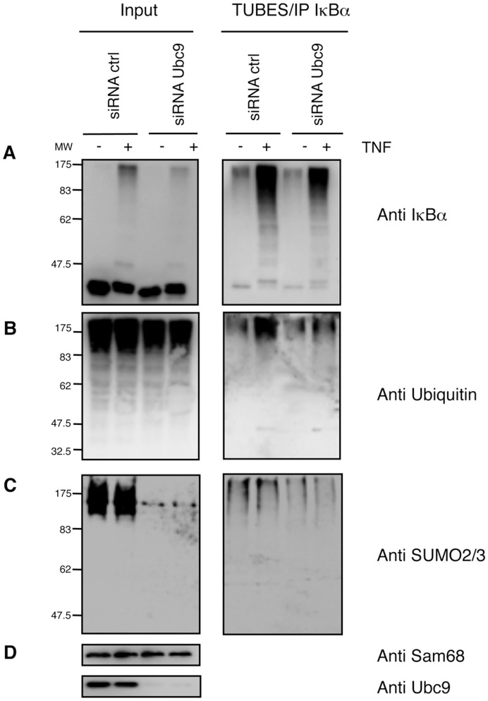
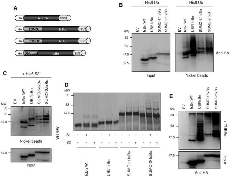
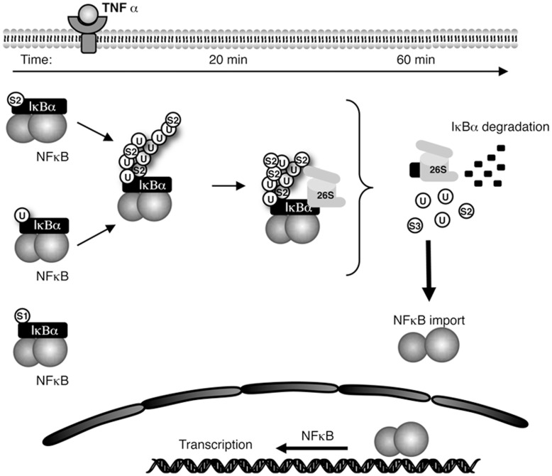
The NF-κB pathway is regulated by SUMOylation at least at three levels: the inhibitory molecule IκBα, the IKK subunit γ/NEMO and the p52 precursor p100. Here we investigate the role of SUMO-2/3 in the degradation of IκBα and activation of NF-κB mediated by TNFα. We found that under conditions of deficient SUMOylation, an important delay in both TNFα-mediated proteolysis of IκBα and NF-κB dependent transcription occurs. In vitro and ex vivo approaches, including the use of ubiquitin-traps (TUBEs), revealed the formation of chains on IκBα containing SUMO-2/3 and ubiquitin after TNFα stimulation. The integration of SUMO-2/3 appears to promote the formation of ubiquitin chains on IκBα after activation of the TNFα signalling pathway. Furthermore, heterologous chains of SUMO-2/3 and ubiquitin promote a more efficient degradation of IκBα by the 26S proteasome in vitro compared to chains of either SUMO-2/3 or ubiquitin alone. Consistently, Ubc9 silencing reduced the capture of IκBα modified with SUMO-ubiquitin hybrid chains that display a defective proteasome-mediated degradation. Thus, hybrid SUMO-2/3-ubiquitin chains increase the susceptibility of modified IκBα to the action of 26S proteasome, contributing to the optimal control of NF-κB activity after TNFα-stimulation.


Conflict of interest statement

Competing Interests: The authors have declared that no competing interests exist.

Figures

Figure 1
Figure 1. SUMOylation contributes to the optimal TNFα-mediated NF-κB activation and degradation of IκBα.
(A) HeLa cells were transfected 72 h with control or Ubc9 siRNA (100 nM). Cells were co-transfected with a NF-κB-luciferase reporter plasmid (3EnhancerConA) and β-galactosidase reporter. Twenty-four hours later cells were stimulated with TNFα (15 ng/ml) as indicated and luciferase and β-galactosidase activities measured as previously described . The graph corresponds to the mean of three independent experiments. (B) HeLa cells were transfected during 72 h with control or Ubc9 siRNA (100 nM) and stimulated with TNFα (15 ng/ml) as indicated. Western-blot analyses were performed with the indicated antibodies.
Figure 2
Figure 2. IκBα is modified by SUMO-2/3 in vitro and ex vivo.
(A) In vitro SUMOylation assay using IκBα WT or mutated on lysines 21 and 22 as substrates. (B) HEK293 cells were transfected with the indicated plasmids, pre-treated or not with MG132 and stimulated or not with TNFα. His6-ubiquitylated or SUMOylated proteins were purified using denaturing conditions and Ni2+ chromatography.
Figure 3
Figure 3. IκBα is modified by ubiquitin chains containing SUMO-2/3.
(A) HEK293 cells were transfected with the indicated plasmids, pre-treated or not with MG132 and stimulated or not with TNFα. His6-ubiquitylated or SUMOylated proteins were purified using denaturing conditions and Ni2+ chromatography. (B) HEK293 cells were transfected with the indicated plasmids at two different concentrations 1 µg or 2 µg of each constructs. Empty vector (EV) was also used to compensate plasmid DNA to final concentration of 2 or 4 µg respectively. Cells were pre-treated with MG132 and stimulated with TNFα during the indicated times. His6-ubiquitylated or SUMOylated proteins were purified using denaturing conditions and Ni2+ chromatography. Captured material was analysed by western-blot with the indicated antibodies. (C) HEK293 cells were transfected with IκBα-SV5 WT or mutated on K21 and K22 in the presence of His6Ubiquitin, His6-SUMO2 and His6-SUMO3, pre-treated with MG132 and stimulated with TNFα. His6-modified proteins were purified using denaturing conditions and Ni2+ chromatography procedure (D) Time-course modification of IκBα after TNFα-stimulation analysed by immunoprecipitations with anti-IgG control, anti-ubiquitin, anti-IκBα and anti-SUMO-2/3 antibodies. Cells were treated with MG132, stimulated with TNFα and lysates were submitted to immunoprecipitation experiments as indicated. Precipitated material was analysed by western-blot with anti-IκBα antibody.
Figure 4
Figure 4. IκBα modified with hybrid SUMO-Ubiquitin chains is captured using Ubiquitin-traps.
(A) HEK293 cells were pre-treated with MG132 and stimulated with TNFα for 20 min. Cells were lysed in a buffer containing TUBE-hHR23A or GST used as a control. GST-captured material was eluted and submitted to IκBα immunoprecipitation. (B) Cells were treated as in (A) and lysed in a buffer containing TUBE-hHR23A. Captured material was eluted and submitted to IκBα or control immunoprecipitations. (C) Cells were treated with MG132 and stimulated with TNFα for the indicated time. Cells were lysed in a buffer containing TUBE-hHR23A. Captured material was eluted and submitted to IκBα immunoprecipitation. (D) Cells were treated as in (C) and lysed in a buffer containing TUBE-hHR23A. Captured material was eluted and submitted to IκBα, ubiquitin or SUMO2/3 immunoprecipitations.
Figure 5
Figure 5. Integration of SUMO molecules into IκBα Ubiquitin chains.
Seventy-two hours after transfection with control or Ubc9 siRNA (100 nM), HeLa cells were pre-treated with MG132, stimulated 20 min with TNFα and lysed in a buffer containing TUBE-hHR23A. TUBE-captured material was eluted and submitted to IκBα immunoprecipitation. Western blot detection with (A) anti-IκBα, (B) anti-ubiquitin, (C) anti-SUMO2/3 and (D) anti-sam68 and anti-Ubc9 antibodies.
Figure 6
Figure 6. SUMO-2 and Ubiquitin promote efficient chain extension on IκBα.
(A) Strategy used to make the different fusions proteins. (B) HEK293 cells were co-transfected with His6-ubiquitin and IκBα fusion-proteins as indicated. Cells were pre-treated with MG132 and stimulated 20 min with TNFα. His6-ubiquitylated proteins were purified using denaturing conditions and Ni2+ chromatography. EV: Empty Vector. (C) HEK293 cells were co-transfected with His6-SUMO-2 and IκBα fusions protein as indicated. Cells were pre-treated with MG132 and stimulated 20 min with TNFα as in A. His6-sumoylated proteins were purified using Ni2+ chromatography procedure. (D) In vitro SUMOylation assay using IκBα WT or fusion proteins as substrates. (E) HEK293 cells were transfected as indicated, pre-treated with MG132 and stimulated 20 min with TNFα. Cells were lysed in a buffer containing 3.5 µM of TUBE hHR23A. TUBE-captured material was eluted and submitted to IκBα immunoprecipitation. EV: Empty Vector.
Figure 7
Figure 7. SUMO-2/3-Ubiquitin chains drive an efficient IκBα degradation by the 26S proteasome.
(A) (B) In vitro ubiquitylation, SUMOylation or mixed assays using IκBα WT (A) or S35 IκBα WT (B) as substrates. Suboptimal conditions of conjugation were used in this assay (see materials and methods). (A) Western blot detection with the indicated antibodies. (B) Detection of radio-labelled material. (C) In vitro ubiquitylation, SUMOylation or mixed assays using S35 IκBα WT as substrate in the presence (+) or absence (-) of 26S proteasome. Saturating conditions of conjugation were used in this assay (see materials and methods). Different Ubiquitin: SUMO-2/SUMO-3 molar ratios were tested as follows: lane 1 = 4∶0/0, lane 2 = 3∶0.5/0.5, lane 3 = 2∶1/1, lane 4 = 1∶1.5/1.5, lane 5∶0:2/2. Detection of radio labelled material. (D) In vitro ubiquitylation, SUMOylation or mixed assays using S35 IκBα WT as substrate in the presence (+) or absence (−) of 26S proteasome. Replicated reactions using saturating conditions and the following ubiquitin: SUMO-2/SUMO-3 ratios: 4∶0/0 for lanes 1 and 2, 0∶2/2 for lanes 3 and 4 and 2∶1/1 for lanes 5 and 6. Phosphorimager quantification of modified forms of S35 IκBα WT in the presence or absence of 26S (n = 5). Standard deviation is indicated in the histograms. (E). Seventy-two hours after transfection with control or Ubc9 siRNA (100 nM), HeLa cells were pre-treated with MG132, stimulated with TNFα and lysed in a buffer containing TUBE-hHR23A. TUBE-captured material was submitted to IκBα immunoprecipitation. After IκBα-IP, extracts were eluted with glycine 200 mM pH2.5, equilibrated at pH 7.5 and submitted to an in vitro proteasome-mediated degradation assay at the indicated times.
Figure 8
Figure 8. Integrated view of the time-dependent contribution of SUMO-2/3 in the formation of ubiquitin chains controlling the proteasomal degradation of IκBα and optimising NF-κB activity.

References

    1. Hay RT (2005) SUMO: a history of modification. Mol Cell 18: 1–12. - PubMed
    1. Hershko A, Ciechanover A (1998) The ubiquitin system. Annu Rev Biochem 67: 425–479. - PubMed
    1. Chen ZJ (2005) Ubiquitin signalling in the NF-kappaB pathway. Nat Cell Biol 7: 758–765. - PMC - PubMed
    1. Rahighi S, Ikeda F, Kawasaki M, Akutsu M, Suzuki N, et al. (2009) Specific recognition of linear ubiquitin chains by NEMO is important for NF-kappaB activation. Cell 136: 1098–1109. - PubMed
    1. Tokunaga F, Sakata S, Saeki Y, Satomi Y, Kirisako T, et al. (2009) Involvement of linear polyubiquitylation of NEMO in NF-kappaB activation. Nat Cell Biol 11: 123–132. - PubMed

Publication types

MeSH terms