

High-glucose-induced endothelial cell injury is inhibited by a Peptide derived from human apolipoprotein E
- PMID: 23284911
- PMCID: PMC3526597
- DOI: 10.1371/journal.pone.0052152
High-glucose-induced endothelial cell injury is inhibited by a Peptide derived from human apolipoprotein E
Abstract
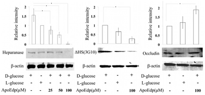
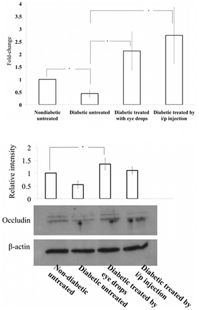
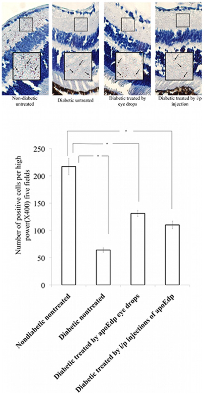
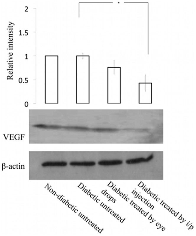
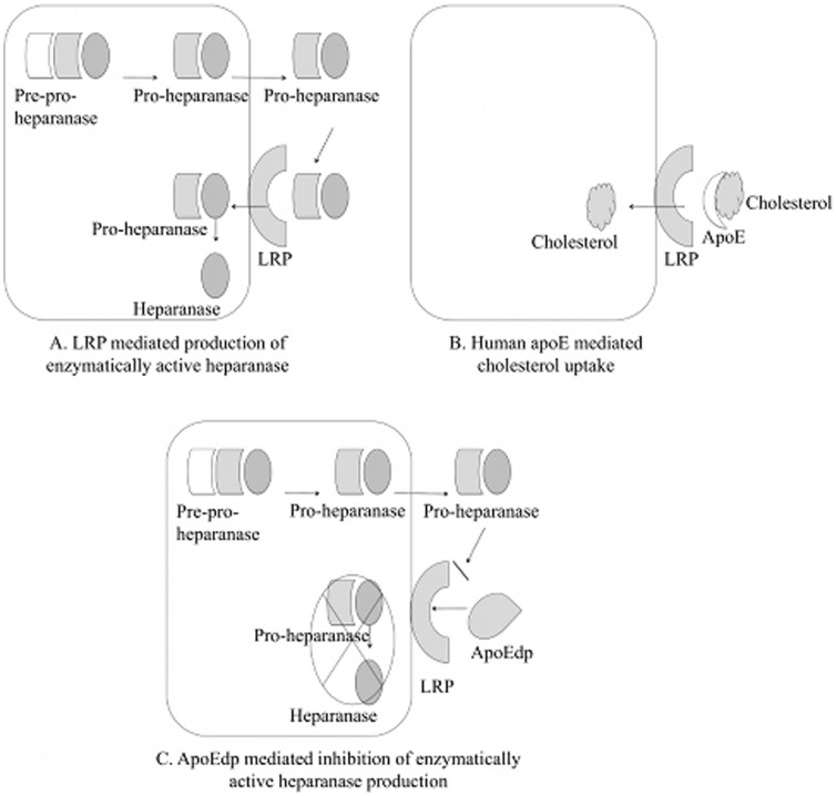
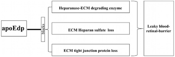
Although the importance of human apolipoprotein E (apoE) in vascular diseases has clearly been established, most of the research on apoE has focused on its role in cholesterol metabolism. In view of the observation that apoE and its functional domains impact extracellular matrix (ECM) remodeling, we hypothesized that apoE could also confer protection against ECM degradation by mechanisms independent of its role in cholesterol and lipoprotein transport. The ECM degrading enzyme, heparanase, is secreted by cells as pro-heparanase that is internalized through low-density lipoprotein (LDL) receptor-related protein-1 (LRP-1) to become enzymatically active. Both apoE and pro-heparanase bind the LRP-1. We further hypothesized that an apoE mimetic peptide (apoEdp) would inhibit the production of active heparanase by blocking LRP-1-mediated uptake of pro-heparanase and thereby decrease degradation of the ECM. To test this hypothesis, we induced the expression of heparanase by incubating human retinal endothelial cells (hRECs) with high glucose (30 mM) for 72 hours. We found that elevated expression of heparanase by high glucose was associated with increased shedding of heparan sulfate (ΔHS) and the tight junction protein occludin. Treatment of hRECs with 100 µM apoEdp in the presence of high glucose significantly reduced the expression of heparanase, shedding of ΔHS, and loss of occludin as detected by Western blot analysis. Either eye drop treatment of 1% apoEdp topically 4 times a day for 14 consecutive days or intraperitoneal injection (40 mg/kg) of apoEdp daily for 14 consecutive days in an in vivo mouse model of streptozotocin-induced diabetes inhibited the loss of tight junction proteins occludin and zona occludin- 1 (ZO-1). These findings imply a functional relationship between apoE and endothelial cell matrix because the deregulation of these molecules can be inhibited by a short peptide derived from the receptor-binding region of apoE. Thus, strategies targeting ECM-degrading enzymes could be therapeutically beneficial for treating diabetic retinopathy.
Conflict of interest statement
Figures
Similar articles
-
LRP-1 Pathway Targeted Inhibition of Vascular Abnormalities in the Retina of Diabetic Mice.Curr Eye Res. 2017 Apr;42(4):640-647. doi: 10.1080/02713683.2016.1203441. Epub 2016 Jul 21. Curr Eye Res. 2017. PMID: 27442082 Free PMC article.
-
Upregulation of heparanase in high-glucose-treated endothelial cells promotes endothelial cell migration and proliferation and correlates with Akt and extracellular-signal-regulated kinase phosphorylation.Mol Vis. 2012;18:1684-95. Epub 2012 Jun 20. Mol Vis. 2012. PMID: 22773906 Free PMC article.
-
Retinal heparanase expression in streptozotocin-induced diabetic rats.Can J Ophthalmol. 2010 Feb;45(1):46-51. doi: 10.3129/i09-200. Can J Ophthalmol. 2010. PMID: 20130710
-
The role of the low-density lipoprotein receptor-related protein 1 (LRP-1) in regulating blood-brain barrier integrity.Rev Neurosci. 2016 Aug 1;27(6):623-34. doi: 10.1515/revneuro-2015-0069. Rev Neurosci. 2016. PMID: 27206317 Review.
-
Heparanase: structure, biological functions, and inhibition by heparin-derived mimetics of heparan sulfate.Curr Pharm Des. 2007;13(20):2057-73. doi: 10.2174/138161207781039742. Curr Pharm Des. 2007. PMID: 17627539 Review.
Cited by
-
Endothelial glycocalyx in retina, hyperglycemia, and diabetic retinopathy.Am J Physiol Cell Physiol. 2023 May 1;324(5):C1061-C1077. doi: 10.1152/ajpcell.00188.2022. Epub 2023 Mar 20. Am J Physiol Cell Physiol. 2023. PMID: 36939202 Free PMC article. Review.
-
Simvastatin alleviates hyperpermeability of glomerular endothelial cells in early-stage diabetic nephropathy by inhibition of RhoA/ROCK1.PLoS One. 2013 Nov 14;8(11):e80009. doi: 10.1371/journal.pone.0080009. eCollection 2013. PLoS One. 2013. PMID: 24244596 Free PMC article.
-
Microvascular destabilization and intricated network of the cytokines in diabetic retinopathy: from the perspective of cellular and molecular components.Cell Biosci. 2024 Jun 27;14(1):85. doi: 10.1186/s13578-024-01269-7. Cell Biosci. 2024. PMID: 38937783 Free PMC article. Review.
-
Protective effects of bestatin in the retina of streptozotocin-induced diabetic mice.Exp Eye Res. 2016 Aug;149:100-106. doi: 10.1016/j.exer.2016.06.016. Epub 2016 Jun 23. Exp Eye Res. 2016. PMID: 27344955 Free PMC article.
-
Altered transendothelial transport of hormones as a contributor to diabetes.Diabetes Metab J. 2014 Apr;38(2):92-9. doi: 10.4093/dmj.2014.38.2.92. Diabetes Metab J. 2014. PMID: 24851202 Free PMC article. Review.
References
-
- Ruderman NB, Williamson JR, Brownlee M (1992) Glucose and diabetic vascular disease. FASEB J 6: 2905–2914. - PubMed
-
- Kamata K, Miyata N, Abiru T, Kasuya Y (1992) Functional changes in vascular smooth muscle and endothelium of arteries during diabetes mellitus. Life Sci 50: 1379–1387. - PubMed
-
- Aiello LP, Avery RL, Arrigg PG, Keyt BA, Jampel HD, et al. (1994) Vascular endothelial growth factor in ocular fluid of patients with diabetic retinopathy and other retinal disorders. N Engl J Med 331: 1480–1487. - PubMed
-
- Muir H (1977) Structure and function of proteoglycans of cartilage and cell-matrix interactions. Soc Gen Physiol Ser 32: 87–99. - PubMed
Publication types
MeSH terms
Substances
Grants and funding
LinkOut - more resources
Full Text Sources
Other Literature Sources
Research Materials
Miscellaneous
