. 2012;7(12):e52208.
doi: 10.1371/journal.pone.0052208. Epub 2012 Dec 20.

Natalizumab exerts direct signaling capacity and supports a pro-inflammatory phenotype in some patients with multiple sclerosis

Affiliations

Natalizumab exerts direct signaling capacity and supports a pro-inflammatory phenotype in some patients with multiple sclerosis

Thomas F Benkert et al. PLoS One. 2012.

Abstract

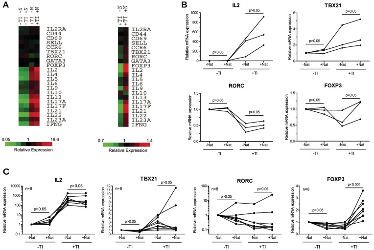
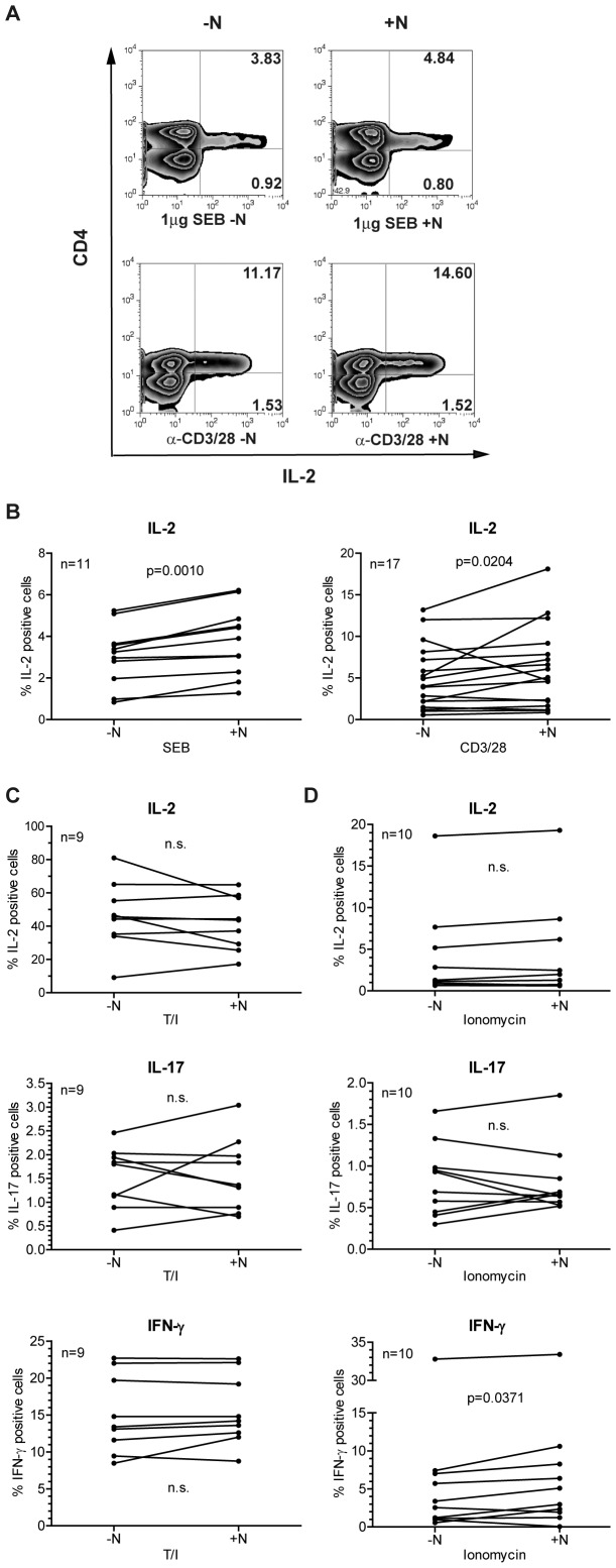
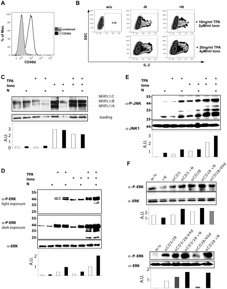
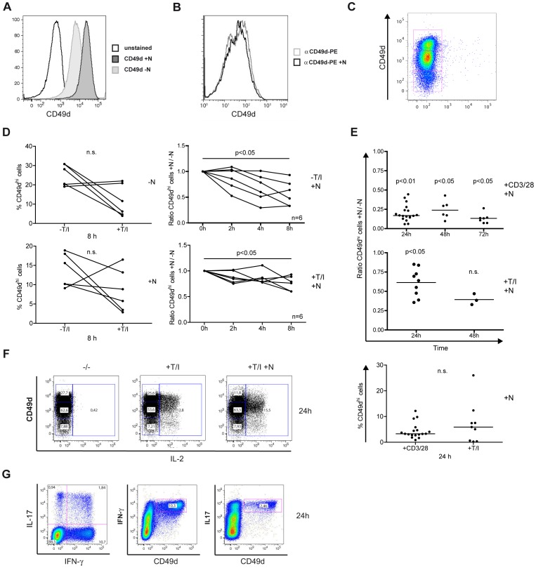
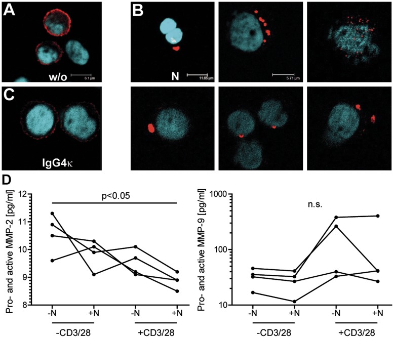
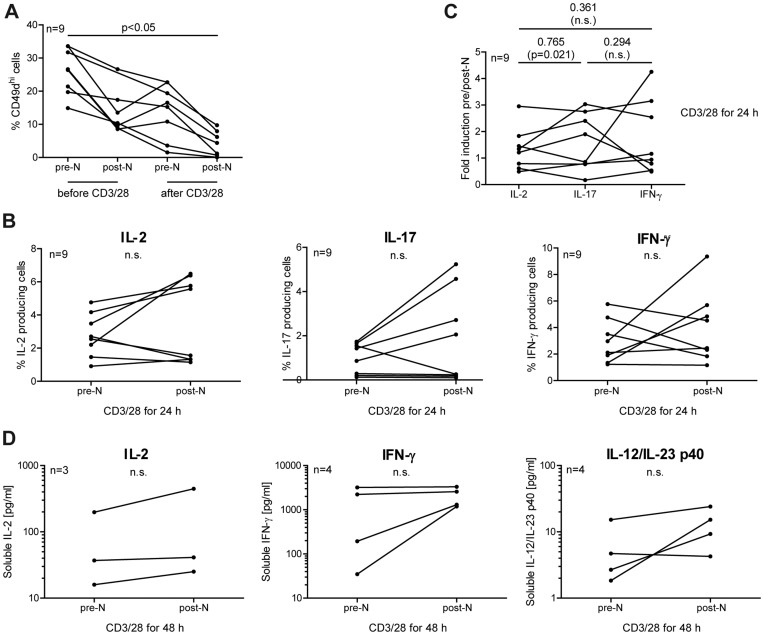
Natalizumab is a recombinant monoclonal antibody raised against integrin alpha-4 (CD49d). It is approved for the treatment of patients with multiple sclerosis (MS), a chronic inflammatory autoimmune disease of the CNS. While having shown high therapeutic efficacy, treatment by natalizumab has been linked to progressive multifocal leukoencephalopathy (PML) as a serious adverse effect. Furthermore, drug cessation sometimes induces rebound disease activity of unknown etiology. Here we investigated whether binding of this adhesion-blocking antibody to T lymphocytes could modulate their phenotype by direct induction of intracellular signaling events. Primary CD4(+) T lymphocytes either from healthy donors and treated with natalizumab in vitro or from MS patients receiving their very first dose of natalizumab were analyzed. Natalizumab induced a mild upregulation of IL-2, IFN-γ and IL-17 expression in activated primary human CD4(+) T cells propagated ex vivo from healthy donors, consistent with a pro-inflammatory costimulatory effect on lymphokine expression. Along with this, natalizumab binding triggered rapid MAPK/ERK phosphorylation. Furthermore, it decreased CD49d surface expression on effector cells within a few hours. Sustained CD49d downregulation could be attributed to integrin internalization and degradation. Importantly, also CD4(+) T cells from some MS patients receiving their very first dose of natalizumab produced more IL-2, IFN-γ and IL-17 already 24 h after infusion. Together these data indicate that in addition to its adhesion-blocking mode of action natalizumab possesses mild direct signaling capacities, which can support a pro-inflammatory phenotype of peripheral blood T lymphocytes. This might explain why a rebound of disease activity or IRIS is observed in some MS patients after natalizumab cessation.


Conflict of interest statement

Competing Interests: M.B. received a travel grant from Biogen Idec, the manufacturer of natalizumab (Tysabri®), which was not related to this manuscript. Furthermore, he received travel grants, speaker’s fees and a research grant from Merck Serono, served on an advisory board for Almirall Hermal GmbH, and received research grants from Sanofi Aventis and Teva Pharmaceutical Industries Ltd. as well as from Novartis. The other authors declare no competing interests. Biogen Idec/Elan had no role in study design, data collection and analysis, decision to publish, or preparation of the manuscript. This does not alter the authors’ adherence to all the PLOS ONE policies on sharing data and materials.

Figures

Figure 1
Figure 1. Top ten genes with natalizumab-driven differential expression levels.
(A) List of genes that were influenced the most by natalizumab in CD4+ T cells, which were restimulated for 8 h. (B) List of genes that were influenced the most by natalizumab in CD4+ T cells, which were not restimulated.
Figure 2
Figure 2. Natalizumab supports a pro-inflammatory gene expression profile.
(A) RNA expression differences of three independent biological replicates are depicted in heat maps for immunologically relevant genes. Cells had not or had been restimulated with T/I (TI). Both groups had been grown with or without natalizumab (N or Nat). (B) qRT-PCR of RNA used for the microarray (triplicates) was analyzed for RNA levels of IL-2 as well as RORγt, T-BET and FOXP3. These three transcription factors are transcribed from the genes RORC, TBX21 and FOXP3, respectively. (C) Independent replication of the qRT-PCR results in 8 different additional donors. Here, CD4+ T cells were stimulated by anti-CD3/28-Dynabeads® for 10 days and restimulated with T/I for 8 h. Dots represent single donors, lines connect intraindividual values. Friedman tests followed by Dunn’s post tests were performed in (B) and (C).
Figure 3
Figure 3. Intracellular protein levels of IL-2, IL-17 and IFN-γ are enhanced by concomitant treatment with natalizumab.
(A, B) PBMC were stimulated by SEB or anti-CD3/28 without or in the presence of natalizumab for 24 h. After surface staining for CD4 and intracellular staining for IL-2, cells were analyzed by FACS. (A) Representative dot blots are given. (B) Single values of samples from n = 11 (SEB) and n = 17 (anti-CD3/28) donors are shown. (C, D) Isolated CD4+ T cells were stimulated with T/I (n = 9) or ionomycin (n = 10) for 8 h. Intracellular FACS stainings for IL-2, IL-17, and IFN-γ were performed, and the differences between cultures with and without natalizumab were calculated. Two-tailed Wilcoxon matched-pairs signed rank tests were performed for statistical analysis in (B, C, and D).
Figure 4
Figure 4. Natalizumab signals via rapid phosphorylation of ERK.
(A) Surface expression of CD49d was verified for Jurkat cells. (B) Intracellular FACS staining for IL-2 revealed increased expression in the presence of natalizumab, when cells were stimulated by T/I (2 concentrations) for 8 h. (C) Western blot analysis of nuclear extracts for NFATc1 after stimulation as indicated for 2 h. (D, E) Immunoblot of proteins extracted from Jurkat cells after stimulation with TPA, ionomycin or both in the presence or absence of natalizumab for 2 h. (D) Detection of pERK and ERK within whole cell extracts. (E) Detection of pJNK and JNK within whole cell extracts. (F) Immunoblot of total proteins extracted from human CD4+ T cells after stimulation with anti-CD3/28– alone or in combination - in the absence or presence of either agonistic anti-CD49d or natalizumab for 2 h. Phosphorylated ERK and total ERK were detected.
Figure 5
Figure 5. Natalizumab reduces CD49d surface expression.
(A) Jurkat cells were treated with natalizumab for 24 h and stained for CD49d expression. (B) A possible interference of natalizumab and the staining antibody for CD49d was evaluated. (C) T/I-activated PBMC incubated with natalizumab for 24 h prior to staining and FACS analyses. Gated on CD4+ T cells, CD49d appeared in three subpopulations. (D) Percentage of CD49dhi cells among all CD4+ cells after stimulation as indicated for 8 h (left column) and normalization of natalizumab-stimulated to natalizumab-unstimulated cells without and with T/I activation for 2, 4 and 8 h (right column). CD49dhi expression was evaluated by FACS. (E) CD4+ T cells were activated with CD3/28 or T/I in the absence or presence of natalizumab for the indicated durations. Ratios of natalizumab-stimulated vs. unstimulated cells were calculated (upper two diagrams). Lower diagram shows percentage of CD49dhi cells among all CD4+ cells after activation with CD3/28 or T/I for 24 h. Same samples as in first columns of upper and middle diagram. (F) CD4+ T cells had been preincubated for 24 h with natalizumab, finally stimulated by T/I for 8 h, stained for CD49d surface and IL-2 intracellular expression and evaluated by FACS. (G) CD4+ T cells were stimulated by T/I for 8 h and stained for intracellular IL-17 and IFN-γ as well as for surface expression of CD49d, analyzed by FACS.
Figure 6
Figure 6. Integrin α-4 is subject to rapid internalization and degradation upon natalizumab binding.
(A–C) Immunocytochemical staining of CD49d and counter-staining of nuclei in PBMC that were either left untreated (A), stimulated with 30 µg/ml natalizumab for 24 h (B) or stimulated with 30 µg/ml IgG4κ isotype control (C). Cells were fixed with 3.7% PFA and permeabilized with 0.1% Triton X-100 before staining. Representative of samples from 4 different donors. (D) PBMC were stimulated with anti-CD3/28 and/or natalizumab as indicated for 24 h. Subsequently, total protein levels of MMP-2 and MMP-9 were determined in cell culture supernatants by commercial ELISA kits. Friedman tests, followed by Dunn’s post tests were performed for statistical analysis.
Figure 7
Figure 7. Natalizumab influences CD4+ T cells from MS patients immediately after the very first infusion.
Pre- and post-natalizumab blood samples were taken from 9 patients with RRMS. (A-C) PBMC were cultivated with plastic-coated anti-CD3/28 for 24 h. (A) Cells were stained for surface expression of CD4 and CD49d before and after in vitro restimulation. (B, C) Cells were stained for intracellular IL-2, IL-17 and IFN-γ concomitantly with staining for surface CD4, and analyzed by FACS. (D) as in A-C, but in vitro restimulation for 48 h. Secretion of interleukins was evaluated by cytometric bead arrays. A two-tailed Kruskal-Wallis test followed by Dunn’s posttest was used in (A) for statistical analysis (no matched pairs test possible due to one missing value). Two-tailed Wilcoxon matched-pairs signed rank tests were performed for statistical analysis in (B) and (D). Spearman correlation coefficients were calculated in (C).

References

    1. Compston A, Coles A (2002) Multiple sclerosis. Lancet 359: 1221–1231. - PubMed
    1. Hohlfeld R, Wekerle H (2001) Immunological update on multiple sclerosis. Curr Opin Neurol 14: 299–304. - PubMed
    1. Korn T (2008) Pathophysiology of multiple sclerosis. J Neurol 255 Suppl 62–6. - PubMed
    1. Benvenuto R, Paroli M, Buttinelli C, Franco A, Barnaba V, et al. (1992) Tumor necrosis factor-alpha and interferon-gamma synthesis by cerebrospinal fluid-derived T cell clones in multiple sclerosis. Ann N Y Acad Sci 650: 341–346. - PubMed
    1. Voskuhl RR, Martin R, Bergman C, Dalal M, Ruddle NH, et al. (1993) T helper 1 (Th1) functional phenotype of human myelin basic protein-specific T lymphocytes. Autoimmunity 15: 137–143. - PubMed

Publication types

MeSH terms