Effects on contralateral muscles after unilateral electrical muscle stimulation and exercise
- PMID: 23284946
- PMCID: PMC3527434
- DOI: 10.1371/journal.pone.0052230
Effects on contralateral muscles after unilateral electrical muscle stimulation and exercise
Abstract
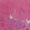
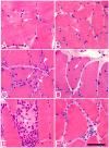
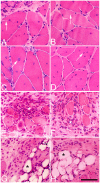
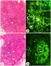
It is well established that unilateral exercise can produce contralateral effects. However, it is unclear whether unilateral exercise that leads to muscle injury and inflammation also affects the homologous contralateral muscles. To test the hypothesis that unilateral muscle injury causes contralateral muscle changes, an experimental rabbit model with unilateral muscle overuse caused by a combination of electrical muscle stimulation and exercise (EMS/E) was used. The soleus and gastrocnemius muscles of both exercised and non-exercised legs were analyzed with enzyme- and immunohistochemical methods after 1, 3 and 6 weeks of repeated EMS/E. After 1 w of unilateral EMS/E there were structural muscle changes such as increased variability in fiber size, fiber splitting, internal myonuclei, necrotic fibers, expression of developmental MyHCs, fibrosis and inflammation in the exercised soleus muscle. Only limited changes were found in the exercised gastrocnemius muscle and in both non-exercised contralateral muscles. After 3 w of EMS/E, muscle fiber changes, presence of developmental MyHCs, inflammation, fibrosis and affections of nerve axons and AChE production were observed bilaterally in both the soleus and gastrocnemius muscles. At 6 w of EMS/E, the severity of these changes significantly increased in the soleus muscles and infiltration of fat was observed bilaterally in both the soleus and the gastrocnemius muscles. The affections of the muscles were in all three experimental groups restricted to focal regions of the muscle samples. We conclude that repetitive unilateral muscle overuse caused by EMS/E overtime leads to both degenerative and regenerative tissue changes and myositis not only in the exercised muscles, but also in the homologous non-exercised muscles of the contralateral leg. Although the mechanism behind the contralateral changes is unclear, we suggest that the nervous system is involved in the cross-transfer effects.
Conflict of interest statement
Figures


Similar articles
-
Unilateral muscle overuse causes bilateral changes in muscle fiber composition and vascular supply.PLoS One. 2014 Dec 29;9(12):e116455. doi: 10.1371/journal.pone.0116455. eCollection 2014. PLoS One. 2014. PMID: 25545800 Free PMC article.
-
Bilateral increase in expression and concentration of tachykinin in a unilateral rabbit muscle overuse model that leads to myositis.BMC Musculoskelet Disord. 2013 Apr 12;14:134. doi: 10.1186/1471-2474-14-134. BMC Musculoskelet Disord. 2013. PMID: 23587295 Free PMC article.
-
Bilateral muscle fiber and nerve influences by TNF-alpha in response to unilateral muscle overuse - studies on TNF receptor expressions.BMC Musculoskelet Disord. 2017 Nov 28;18(1):498. doi: 10.1186/s12891-017-1796-6. BMC Musculoskelet Disord. 2017. PMID: 29183282 Free PMC article.
-
Contributions to the understanding of gait control.Dan Med J. 2014 Apr;61(4):B4823. Dan Med J. 2014. PMID: 24814597 Review.
-
Muscle mechanics: adaptations with exercise-training.Exerc Sport Sci Rev. 1996;24:427-73. Exerc Sport Sci Rev. 1996. PMID: 8744258 Review.
Cited by
-
Marked Effects of Tachykinin in Myositis Both in the Experimental Side and Contralaterally: Studies on NK-1 Receptor Expressions in an Animal Model.ISRN Inflamm. 2013 Jan 29;2013:907821. doi: 10.1155/2013/907821. eCollection 2013. ISRN Inflamm. 2013. PMID: 24049666 Free PMC article.
-
Electrical Impedance Myography to Detect the Effects of Electrical Muscle Stimulation in Wild Type and Mdx Mice.PLoS One. 2016 Mar 17;11(3):e0151415. doi: 10.1371/journal.pone.0151415. eCollection 2016. PLoS One. 2016. PMID: 26986564 Free PMC article.
-
Multiple Applications of Different Exercise Modalities with Rodents.Oxid Med Cell Longev. 2021 Nov 25;2021:3898710. doi: 10.1155/2021/3898710. eCollection 2021. Oxid Med Cell Longev. 2021. PMID: 34868454 Free PMC article. Review.
-
Inhibitors of endopeptidase and angiotensin-converting enzyme lead to an amplification of the morphological changes and an upregulation of the substance P system in a muscle overuse model.BMC Musculoskelet Disord. 2014 Apr 11;15:126. doi: 10.1186/1471-2474-15-126. BMC Musculoskelet Disord. 2014. PMID: 24725470 Free PMC article.
-
Adipose stem cells enhance myoblast proliferation via acetylcholine and extracellular signal-regulated kinase 1/2 signaling.Muscle Nerve. 2018 Feb;57(2):305-311. doi: 10.1002/mus.25741. Epub 2017 Aug 13. Muscle Nerve. 2018. PMID: 28686790 Free PMC article.
References
-
- Koltzenburg M, Wall PD, McMahon SB (1999) Does the right side know what the left is doing? Trends Neurosci 22: 122–127. - PubMed
-
- Zhou S (2000) Chronic neural adaptations to unilateral exercise: mechanisms of cross education. Exerc Sport Sci Rev 28: 177–184. - PubMed
-
- Munn J, Herbert RD, Gandevia SC (2004) Contralateral effects of unilateral resistance training: a meta-analysis. J Appl Physiol 96: 1861–1866. - PubMed
-
- Carroll TJ, Herbert RD, Munn J, Lee M, Gandevia SC (2006) Contralateral effects of unilateral strength training: evidence and possible mechanisms. J Appl Physiol 101: 1514–1522. - PubMed
-
- Bezerra P, Zhou S, Crowley Z, Brooks L, Hooper A (2009) Effects of unilateral electromyostimulation superimposed on voluntary training on strength and cross-sectional area. Muscle Nerve 40: 430–437. - PubMed
Publication types
MeSH terms
LinkOut - more resources
Full Text Sources

