. 2012;7(12):e52234.
doi: 10.1371/journal.pone.0052234. Epub 2012 Dec 20.

In vivo human left-to-right ventricular differences in rate adaptation transiently increase pro-arrhythmic risk following rate acceleration

Affiliations

In vivo human left-to-right ventricular differences in rate adaptation transiently increase pro-arrhythmic risk following rate acceleration

Alfonso Bueno-Orovio et al. PLoS One. 2012.

Abstract

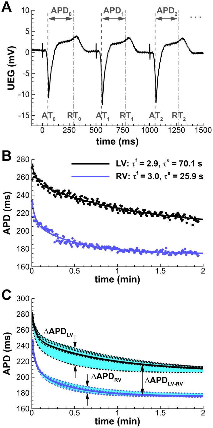
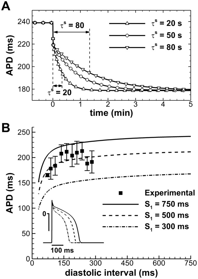
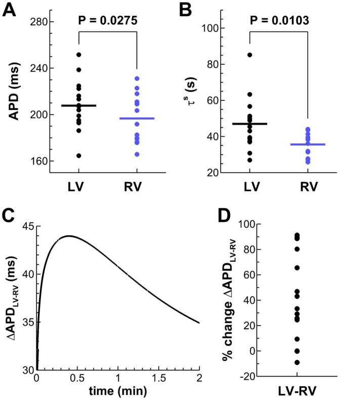
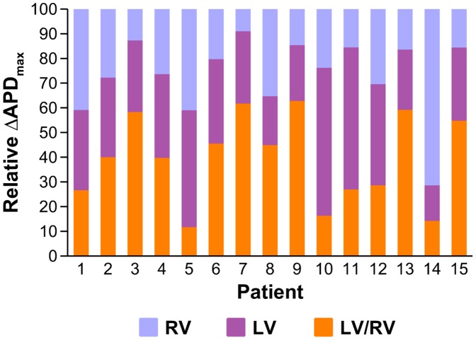
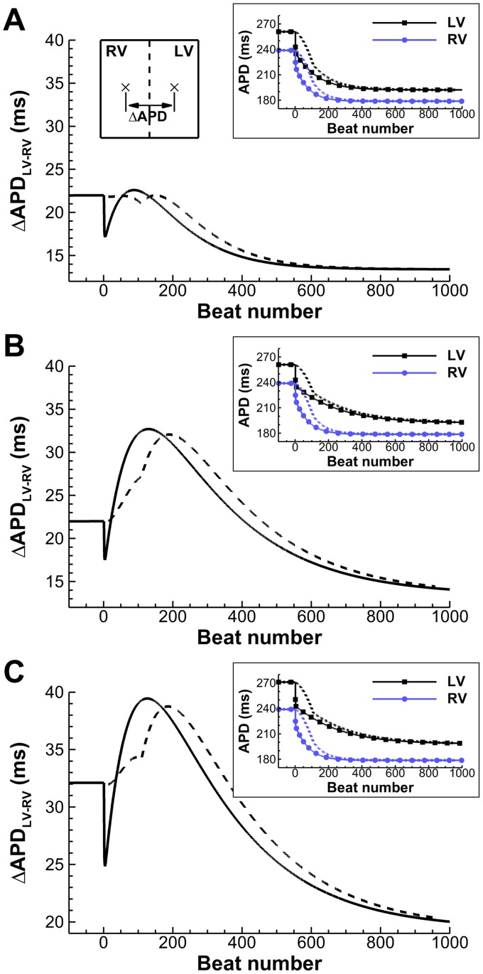
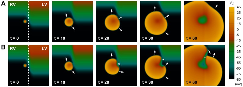
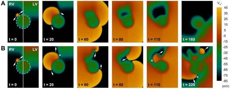
Left-to-right ventricular (LV/RV) differences in repolarization have been implicated in lethal arrhythmias in animal models. Our goal is to quantify LV/RV differences in action potential duration (APD) and APD rate adaptation and their contribution to arrhythmogenic substrates in the in vivo human heart using combined in vivo and in silico studies. Electrograms were acquired from 10 LV and 10 RV endocardial sites in 15 patients with normal ventricles. APD and APD adaptation were measured during an increase in heart rate. Analysis of in vivo electrograms revealed longer APD in LV than RV (207.8 ± 21.5 vs 196.7 ± 20.1 ms; P<0.05), and slower APD adaptation in LV than RV (time constant τ(s) =47.0 ± 14.3 vs 35.6 ± 6.5 s; P<0.05). Following rate acceleration, LV/RV APD dispersion experienced an increase of up to 91% in 12 patients, showing a strong correlation (r(2) =0.90) with both initial dispersion and LV/RV difference in slow adaptation. Pro-arrhythmic implications of measured LV/RV functional differences were studied using in silico simulations. Results show that LV/RV APD and APD adaptation heterogeneities promote unidirectional block following rate acceleration, albeit being insufficient for establishment of reentry in normal hearts. However, in the presence of an ischemic region at the LV/RV junction, LV/RV heterogeneity in APD and APD rate adaptation promotes reentrant activity and its degeneration into fibrillatory activity. Our results suggest that LV/RV heterogeneities in APD adaptation cause a transient increase in APD dispersion in the human ventricles following rate acceleration, which promotes unidirectional block and wave-break at the LV/RV junction, and may potentiate the arrhythmogenic substrate, particularly in patients with ischemic heart disease.


Conflict of interest statement

Competing Interests: The authors have declared that no competing interests exist.

Figures

Figure 1
Figure 1. Quantification of intra- and interventricular APD dispersion (ΔAPD) from unipolar electrocardiograms.
A: Automatic detection of activation and recovery times (AT/RT), and reconstruction of the APD series. The initiation of programmed pacing coincides here with t = 0. B: Estimation of fast (τf) and slow (τs) adaptation time constants by fitting the APD series to a double exponential decay (Patient 3, representative LV/RV mid-ventricular locations). C: Intraventricular ΔAPD is measured as the difference between the longest and shortest APD during adaptation (shaded areas). Solid lines indicate average LV/RV adaptations.
Figure 2
Figure 2. Electrophysiological properties of the human AP model.
A: APD adaptation after a sustained change in rate from normal to fast pacing (750 to 400 ms), for different slow APD adaptation time constants. B: S1–S2 APD restitution at different S1 cycle lengths. Aggregated experimental restitution data at a CL = 500 ms is shown for comparison. Inset shows steady-state APs at the indicated CLs.
Figure 3
Figure 3. Functional LV/RV differences in the in vivo human heart.
A,B: Unipolar electrograms revealed longer steady-state APDs at the study CL and slower APD adaptation dynamics in LV compared to RV. C: Transient increased LV/RV APD dispersion following rate acceleration in a representative patient (Patient 3). D: Percent of ΔAPDLV-RV increase following rate acceleration for all patients of the study.
Figure 4
Figure 4. Comparison of maximum interventricular ΔAPDLV-RV with intraventricular ΔAPDLV and ΔAPDRV for all patients in the study.
Figure 5
Figure 5. Time evolution of ΔAPDLV-RV due to LV/RV differences in APD adaptation, after a sustained (solid) or a gradual (dashed) change in pacing rate (750 to 400 ms). A:
Transient patterns of small ΔAPDLV-RV develop under average conditions of slow APD adaptation. B: Conditions of protracted slow APD adaptation increase maximum amplitude and time window of the transient ΔAPDLV-RV pattern. C: Conditions of larger ΔAPDLV-RV at normal rate translate into a vertical shift of the transient pattern. Insets show LV/RV APD adaptation in each of the cases, for both stimulation protocols.
Figure 6
Figure 6. Development of unidirectional block due to transient patterns of interventricular APD dispersion.
A: Under average conditions of slow APD adaptation (scenario A), the transient APD dispersion between both ventricles only affects wavefront propagation partially, and the ectopic stimulation excites the whole tissue as a regular beat. B: For conditions of protracted slow APD adaptation (scenario B), a larger interventricular APD dispersion is able to produce unidirectional block (t = 20, marked by an asterisk), leading to the initiation of reentry (t = 30), that subsequently develops in the tissue (t = 60). Colorbar denotes transmembrane potential (mV); times indicated since initiation of ectopic stimulation (ms).
Figure 7
Figure 7. Interaction of transient patterns of interventricular APD dispersion with structural defects of the tissue.
The dashed line indicates an inexcitable region in the LV/RV junction. A: Under average conditions of slow APD adaptation (scenario A), interventricular APD dispersion is not able to produce conduction block, and the extra-stimulus proceeds circumventing the inexcitable area. B: For conditions of protracted slow APD adaptation (scenario B), the top part of the extra activation now finds a region of unidirectional block due to a larger APD dispersion (t = 20, marked by an asterisk). The wavefront therefore moves upwards, eventually developing into a reentrant wave (t = 60). Since the bottom part of the excitation has been circumventing the obstacle, the top reentrant wave can now proceed in the tissue (t = 80), finding new areas of conduction block (t = 110), and finally producing wave-break (t = 220). Figure annotation as in Figure 6.

References

    1. Kuo CS, Munakata K, Reddy CP, Surawicz B (1983) Characteristics and possible mechanisms of ventricular arrhythmia dependent on the dispersion of action potential durations. Circulation 67: 1356–1357. - PubMed
    1. Morgan JM, Cunningham D, Rowland E (1992) Dispersion of monophasic action potential duration: demonstrable in humans after premature ventricular extrastimulation but not in steady state. J Am Coll Cardiol 19: 1244–1253. - PubMed
    1. Yuan S, Wohlfart B, Olsson SB, Blomstrom-Lundqvist C (1995) The dispersion of repolarization in patients with ventricular tachycardia. A study using simultaneous monophasic action potential recordings from two sites in the right ventricle. Eur Heart J 16: 68–76. - PubMed
    1. Clayton RH, Taggart P (2005) Regional differences in APD restitution can initiate wavebreak and re-entry in cardiac tissue: a computational study. Biomed Eng Online 20: 4–54. - PMC - PubMed
    1. Nash MP, Bradley CP, Sutton PM, Clayton RH, Kallis P, et al. (2006) Whole heart action potential duration restitution properties in cardiac patients: a combined clinical and modelling study. Exp Physiol 91: 339–354. - PubMed

Publication types